Anaemia can be defined as decreased haemoglobin counts or reduced red blood cell counts or reduced oxygen carrying capacity of blood, due to “loss of” or “abnormality of” red blood cells or haemoglobin.
Normal Heamoglobin Counts
6 months to 5 years of age > 11g/dl
5 years to 12 years of age > 11.5g/dl
12years to 16 years of age > 12g/dl
Adult Females (non-pregnant) > 12g/dl
Adult Females (pregnant) > 11gm/dl
Adult Males > 13g/dl
CAUSES OF ANAEMIA
Blood losss
Excessive Red Blood Cell destruction
Heamoglobinopathies
Hypovitaminosis B12
Hypoferremia
Anaemia of Chronic diseases
Autoimmune haemolytic anaemia
Inflamatory bowel diseases
Hypervolemia or water retention due to sodium or other salts.
Genetic hereditary conditions like Thalasemia
Certain cancers
Kidney diseases
Reduced erythropoetin production
Excessive RBC destruction
Impaired RBC production
Certain infections like malaria which causes RBC destruction.
Certain drugs which causes RBC destruction eg. Quinine causes chinchonism.
Bone Marrow lesions and pathologies
Etc.
CLASSIFICATION OF ANAEMIA
There are many types of anaemias. It can be broadly classified into 7 categories depending upon their causes
Anaemia due to
Blood Loss
Hemolysis
Impaired or abnormal Erythropoesis
Hypervolemia
Chronic Diseases
Nutritional deficiency
Based on RBC morphology it can be classified into 3 groups
Microcytic
Macrocytic
Normocytic
FEW COMMON and RARE TYPES OF ANAEMIA
Iron Deficiency Anaemia
Aplastic Anaemia
Megaloblastic Anaemia
Pernicious Anaemia
Sideroblastic Anaemia
Autoimmune Hemolytic Anaemia
Myelodysplastic Syndrome
Thalasemia
Fanconi Anaemia
Congenital Dyserythropoetic Anaemia
Daimond-Blacfan Anaemia
Myelopthisis
Anaemia of Prematurity
Erythroblastopenia or Pure Red Cell Aplasia
Hereditary Spherocytosis
Hereditary Elliptocytosis
SYMPTOMS
Weakness
Lethargy
In children it affects growth in general
Somnolence, Drowziness in day time
Disturbed sleep at night
Pallor, general pale appearance of skin, mucous membranes and eyes.
Dyspnoea on Exertion.
Reduced Immunity, tendency to catch infections and slow recovery and healing.
Bodyaches
Cyanosis in severe cases
Palpitations
Tachycardia
Low blood pressure
Chest pain
Depression
Craving for indigestible things , PICA
Cold clammy extremities
Oedematous swelling of extremities, dependent oedema
Angina or cardiac failure in severe cases
Will impact general growth and repair of all the vital organs and tissue of the body.
HOMEOPATHIC MEDICINES FOR ANAEMIA
Depending upon the cause of anaemia and general constitution of the patient, one of the following medicines may be called for duty by a homeopathic physician.
Septic Arthritis is invasion of joint by micro-organisms causing inflamation, it is also called infection of joint or Arthritis due to infection or infective arthritis.
CAUSES
VECTORS
Septic Arthritis can be caused due to infection of any of the following vectors
Bacteria
Virus
Fungus
Parasite
Most Common Organisms Known to Cause Septic Arthritis.
Usually its not common to get joint infection unless the immunity of a person is weak or there is history of surgery, prosthesis, deep tissue infections etc.
Usually single joint is infected, less frequently more than one joints are involved.
Most commonly involved joint is knee joint and other freqently involved joints are hip, spine, shoulder, wrist, elbow, sacroilliac and sternoclavicular joints.
Sudden onset of symptoms, fast progression of disease.
Swelling, redness with increased local Heat and pain around joint. Swelling and redness is comparatively much more than other types of arthritis.
Fever with or without chills and headache.
Cant move joint due to severe pain.
Pain aggravated in slightest motion or jarring, pain is ususally aching type with stitching, stinging and pulsating.
DIAGNOSIS OF SEPTIC ARTHRITIS
Athrocentesis – microorganisms usually found on culture, WBC count above 50,000-1,00,000/cubic mm, neutrophils more than 90%, lactate count more than 10mmol/l.
CBC – increased wbc
Blood culture positive for micro-organism
ESR – elevated
CRP – elevated
Procalcitonin – elevated
NAAT – to rule out gonorehoea
Ultrasound – joint effusion
CT scan – Region involved type and extent of damage
MRI – Region involved type and extent of damage
HOMOEOPATHIC MEDICINES FOR SEPTIC ARTHRITIS
Septic Arthritis is a medical emergeny case and requires immediate medical intervention, any delay in treatment can cause damage and complete destruction of joint with in hours to days.
Acute fast acting Homeopathic Medicines are selected with special affinity to joints and pathogenesis that of sudden, severe, acute inflamation with much swelling redness. Below is the list of indicated homeopathic medicines that I prefer in case of infective arthritis.
CARPEL TUNNEL SYNDROME CTS is a condition where in the compression symptoms arise due to bundle compression of structures that pass within carpel tunnel affecting the the Median Nerve.
WHAT IS CARPEL TUNNEL?
CARPEL TUNNEL is normal anatomical structure in our body found in every normal individual. It is formed by carpel bones forming a groove as its floor on dorsal side of hand and flexor retinaculum forming the flat roof of carpel tunnel on palmar side of hand.
It provides attachment and passage at the wrist level for the structures to pass through arm to hand. Muscles related to flexing movements of finger pass through this tunnel alomg with median nerve
Structures Passing Through Carpel Tunnel
Flexor Digitalis Superficialis – 4 tendons
Flexor Digitalis Profundus – 4 tendons
Flexor Pollicis Longus – 1 tendon
Median Nerve
Flexor Carpi Radialis not exactly pass within the carpel tunnel but it traverse through Flexor Retinaculum that is forming the roof of carpel tunnel.
CAUSES OF CARPEL TUNNEL SYNDROME
Anything that exerts pressure to median nerve giving rise to nerve compression symptoms cause carpel tunnel syndrome. In most cases the cause remains obscure and are idiopathic. Any inflamatory process or water retention or metabolites deposition within tissue may cause increase in volume of structures and total content within the carpel tunnel and will cause in pressure of whole bundle which may cause Carpel tunnel due to increased bundle pressure, similarly the adjecent structures to carpel tunnel if inflamed or injured or any overgrowth of it, may exert pressure on carpel tunnel and median nerve within giving rise to carpel tunnel syndrome. Mechanical reasons like wear-tear and injuries are mostly regarded as major factors with ageing and certain genetic, structural and physiological anomalies are known to increase the risk of Carpel Tunnel Syndrome.
Age and Gender
Age – Ageing plays a major role in development of CTS its more commonly found in age above 40yrs
Gender – Female sex is more prone to this condition compared to male.
Mechanical
OCCUPATIONAL – Work related frequent repetitive forceful pressures jerk and vibrations on hand on regular basis where in there is no sufficient time given to repair the wear and tear and the damage tend to accumulate over time.
POSTURAL HABBITS such that it exerts unusual strain on hand and wrist on regular basis.
INJURIES to wrist distal part of forearm and wrist. Fractures involving radius ulna carpel and metacarpel bones like Colle’s Fracture, Boxer’s Fracture etc.
TRIGGER FINGER or TRIGGER THUMBis a condition where in joints of one or more of the digits(finger/thumb) gets stucked up/locked up at certain postion which is difficult to move and moving it with force may cause popping or clicking sound with pain.
Though it can present in any sex and at any age, it is more commonly found in females around age of 50-60 yrs.
CAUSES OF TRIGGER FINGER/THUMB
Its is also termed as digital Stenosing Tenosynovitis, although there is no predominant inflamation to Tendosynovium but inflamation is found in Tendon Sheath. And also its not comfirmed that inflamatory process has any primary role in its development
Though exact cause behind trigger finger/thumb is not known but the risk factors that tend to increase the incidence of this condition are identified
Over straining and over use of hand and fingers espescially activities involving prolonged forceful flexion of digits(fingers/thumb) may be occupational or habbitual routine activities.
Frequent injuries – occupational, accidental or even injuries of planned surgery of hand especially ofter surgery for carpel tunnel syndrome
Though it may involve any digit(thumb/finger) Index finger and Thumb are more frequently involved. One or more digits may be involved.
Patient presents with stucked up digit at certain position. It may be at any level from flexed to extended position usually found at semiflexed position.
This locking up may be persistent for a prolonged period of time or may be momentary and recurrent.
Aggravations are more commonly experienced at night, especially while holding heavy article with hand or while gripping or applying pressure with fingers or hand.
On moving and or forcefully unlocking the stucked finger is bit painful and causes clicking and popping sound
In severe cases the finger may be persistently locked for prolonged period of time with constant pain which may also extent to whole hand and wrist.
DIAGNOSIS OF TRIGGER FINGER/THUMB
Diagnosis of trigger finger/thumb is based on clinical symptomatology of the patient where in inflamation or involvement of tendon sheath of flexors is confirmed and excluding probability of other condition like
Reactive Arthritis was also called Reiter’s Arthritis is RF-negative and HLA-B27 Linked Imflamatory oligoarthritis typical with Enthesitis, accompanied with Inflamatory occular and/or inflamatory genitourinary and other systemic manifestation usually post gastrointestinal or genitourinary infection.
During world war one and two many cases emerged with the Triad of Symptoms viz. Inflamation of Joints, Inflamation of eyes and Inflamation of Uretha. Which drew attention of medical community due to common presentation in many giving it some syndrome like picture. On further investigations it was found out that most of them were exposed to urogenital or Gastro-intestinal infection 1-4 weeks prior to onset of this Triad of Symptoms. This was initially termed as “Fessenger-Leroy-Reiter’s Syndrome” or simply “Reiter’s Syndrome”. But as the physician Hans Conard Julius Reiter was involved in attrocities and war crimes with Hitler, so his name was removed and later renamed and termed as “Reactive Arthritis”.
EPIDEMIOLOGY OF REACTIVE ARTHRITIS
AGE – It more frequently affects age group of 20-40 years.
SEX – It is more common in Males then in Females.
ETHNICITY – Due to its association with HLA-B27 it is frequently found in white race compared to dark race as comparatively HLA-B27 occurs more commonly in white population.
RISK FACTOR – Person with HIV positive status are more prone to develop reactive arthritis.
SIGNS AND SYMPTOMS OF REACTIVE ARTHRITIS
The onset of symptoms of Reactive Arthritis typically starts 4-35 days after an initial infection of gastro-intestinal system or genito-urinary system.
TRIAD OF REACTIVE ARTHRITIS
Reactive arthritis in most of the cases presents where patient cant – SEE, PEE, climb the TREE! due to following Classical Triad of Symptoms of reactive arthritis
i) OLIGOARTHRITIS
Oligoarthritis involving less than five joints. It may frequently involve knee and sacroilliac joint as well. May present itself in additive pattern where it starts with one joint and add another joints subsequently or it may be migratory in pattern where the set of inflamed joints keep changing by addition and simultaneous substraction of joints involved.
ii) NON-GONOCOCCAL GENITOURINARY INFLAMATION
Inflamation of genitourinary system classically presents itself at the onset of the disease. Not always but in many its typically after initial sexual exposure. It presents as frequent burning micturation, uritheritis, prostatitis, balanitis in men and salpingitis, vulvitis and vaginitis in women.
iii) OCCULAR INFLAMATION
Occular Inflamation may present itself as mild conjunctivitis or uveitis in 75% of cases with gastrointestinal origin and 50% of cases with urogenital involvement. patients have intermittent irritation in eyes with blurred vision typically commences at onset of disease.
OTHER SYMPTOMS
Few patients also presents with peculiar symptom which is specific to reactive arthritis, its Keratoderma Blenorrhagica which are small hard nodule commonly appear on soles occasionally on palms and rarely on other parts of body subcutaneous nodules are not incluced. Even in absence of above mentioned triad of symptom the presence of Keratoderma Blenorrhagica is diagnostic for reactive arthritis.
In reactive arthritis; typical to HLA B27 related immunological reactions; involves Entheses that is where skeletal muscles attaches with bones through tendons, where it causes Enthesitis and tendon inflamation especially the tendo-achilles and also fascia in particular Plantar Tendinitis.
Occasionally patients also suffer from dactilitis giving finger sausage-like apperance “sausage finger” due to inflamation.
Mucocutaneous involvement presents as ulcerative or non ulcerative stomatitis, apthous ulcers and geographic tongue are also seen as presentation of this disease
Cardiac involvement causing pericarditis and aortic regurgitation in cases which do no recover soon or if its recurring or progressive.
Gastrointestinal manifestation like pain and cramps with frequent semiformed stools with mucous and insome cases blood due to inflamation and ulcceration in gastrointestinal tract.
Most of the cases of Reactive Arthritis recover within six months, in many cases it keeps comming back time and again and in few it becomes chronic and progressive which may increase risk of severe complications.
COMPLICATIONS OF REACTIVE ARTHRITIS
In chronic progressive and recurring cases the patient may develop following complications
Reactive Arthritis is is HLA B27 linked inflamatory arthritis and enthesitis preceeded by a spell of infection either of genito-urinary system or gastro-intestinal system by following commonly involved organisms
GENITO-URINARY INFECTIONS ASSOCIATED WITH REACTIVE ARTHRITIS
Chlamydia Trachomatis
Ureaplasma Urealyticum
GASTRO-INTESTINAL INFECTIONS ASSOCIATED WITH REACTIVE ARTHRITIS
Salmonella Spp.
Shigella Spp.
Campylobacter Spp.
Yersinia Spp.
4-35 days after the spell of urethritis or food poisoning by above mentioned organisms the symptoms of reactive arthritis sets in, where the synovial fluid has negative culture ans is free from infection and but the HLA B27 linked inflamation is thought to be triggered due to
Autoimmune reaction due to cross reactivity of micro-organism antigen with joint tissue or
Micro-organism antigenic components that may have settled in joint tissue.
DIAGNOSIS OF REACTIVE ARTHRITIS
Clinically the Reactive Arthritis can be diagnosed with help of Sensitivity and Specificity Guidlines laid down by American College of Rheumatology, for clinical diagnosis with given set of presenting symptom, its as follows
Arthritis > 1 month with Urethritis and/or cervicitis has sensitivity of 84.3% and specificity of 98.2%.
Arthritis > 1 month with Urethritis or Cervicitis or bilateral Conjunctivitis has Sensitivity of 85.5% Specificity of 96.4%.
Arthritis, Urethritis and Conjunctivitis has Sensitivity of 50.8% and sensitivity of 98.8%.
Arthritis > 1 month, Conjunctivitis and Urethritis has Sensitivity 48.2% and Specificity of 98.2%.
Patients falling in above criteria or those showing just Keratoderma Blenorrhagica without any other symptoms and other suspected cases can be sent for following test for further evaluation.
HLA B27 testing
Urine routine and culture
STOOL Routine and culture
Throat swab culture
Cervix and Urethral swab culture
Erythrocytes Sedimentation Rate
C-Reactive Protein Test
HOMEOPATHIC TREATMENT FOR REACTIVE ARTHRITIS
Being an immune mediated systemic reaction that too the one that is triggered with different causative agents and even to same agents different individuals will respond differently.
Though they may have same set of general symptoms like the classical triad of reactive arthritis but intensity of each of the symptom of triad will differ in each individual,
Now this is where the homeopathic individualisation process starts. In Homeopathy we believe that though majority of human genome is the same but the minor variations in gene and the epigenome make the whole lot of difference in various characteristerics of each individual, similarly their immune reaction also varies, so every person should have individualised medicine.
Homeopathic Treatment is based on symptom similarity and individualisation of case based on peculiar symptoms based on which the case is individualised and medicine is selected.
Alternatively as per Homeopathic principle of Genus Epidemicus or pathology based symptomatology there can be disease specific homeopathic medicine derived from common symptomatic representation of a disease condition in a group of population.
Now this can not be the most similimum homeopathic prescription but roughly it can hit the disease condition within an indivudual though not accurate but will yeild some results in most of the cases.
To yield best homeopathic results there can be no generalised common approach for all cases.
But still if we have to attempt common standardised pathology based approach then to give some guidelines on homeopathic approach towards cases of reactive arthritis I have attempted following rough guidelines which may help to give some vision in approach towards such cases.
Its seen that in few case it begins after gastro-intestinal infection and in some case post genito-urinary infection. So this will further guide determining “morbid cause” behind the disease directing us in homoeopathic similimum medicine selection.
Now reactive Arthritis shows a triad of symptom in most of the cases. So this triad helps us to reach to group of medicines with such combination of symptoms.
Intensity, occurance of symptoms and its sequence in triad differs in each individuals. For example
In some person urogenital symptoms may be more severe compared to occular symptoms or arthritis symptoms, where as in others arthritis and ocular symptoms would be more severe than urogenital symptoms.
Some may not have occurence of conjunctivitis
In some all three triad occur at a time where as in some patients it may occur gradually one after another in different sequence.
All this helps us find out the “seat of disease” in an individual and its degree of affinity towards various organs which can be related to homeopathic medicines during selection process.
Also similarly symptoms of occular involvement and urogenital involvement should be take in to account in absolute detail. This further helps refine and classify the patient and the respective medicines to be repertorised.
Which other systems and organs are involved like mucous membranes, skin, heart, kidney etc and what type of pathology they are showing like tissue destruction or just inflamation and functional disturbance or tissue lysis with regenerated and degenerative changes this will help to decide what “type of miasm” is underlying wether its psoric, syphillitic or psychotic type pathology.
Certain symptom are very “peculiar” for the disease and occurs in few individuals like Keratoderma Blenorrhagica eruption, now location of this eruption will further help individualise the case.
Enthesitis – Inflamation of tendo-achilles and plantar fascitis is “very specific” to the disease but does not occur in all individuals, so if plantar fascitis or inflamation of tendo-achilles if occurs in someine with this disease then it helps further in individualisation of during homeopathic medicine selection.
Other than this the general health and family background should be noted to derive constitutional types and association of HLA B27 in 75% of this individual further helps in individualisation and homeopathic medicine selection.
COMMONLY USED HOMEOPATHIC MEDICINES FOR REACTIVE ARTHRITIS
Frozen Shoulder also called Adhesive Capsulitis of shoulder joint is painful stiffness limiting range of motion of shoulder joint caused due to inflammation related pathophysiology in capsule of shoulder joint.
CAUSES AND RISK FACTORS OF FROZEN SHOULDER
CAUSE OF FROZEN SHOULDER
Exact cause of Frozen Shoulder remains unclear but there are many factors that increases the risk and lead to frozen shoulder
RISK FACTORS FOR FROZEN SHOULDER
GENDER
Frozen shoulder is more common in females than in males
AGE
Person above 40yrs of age are more prone to this condition
INJURIES
Injury to shoulder joint involving any of its companent especially gleno-humeral joint capsule or rotator cuff.
Sports persons typically develop calcifications of tendons around shoulder joint and also in joint structures due to repeated injury and result of prolonged inflamation such persons are at high risk of developing frozen shoulder.
REDUCED MOBILITY
Injuries to arm and neck may indirectly affect shoulder joint, due lack of motion in general of that region as a whole and changes pattern of gait and movements of that region.
Immobility or reduced mobility post surgerr with general debility and delayed recovery.
Immobility post stroke.
MUSCULO-SKELETAL DEFECTS
Certain congenital structural anomaly of Musculoskeletal framework which has influence on movement, gait and weight distribution pattern on shoulder joint in abnormal pattern, which may put the person at risk of develooing Frozen Shoulder.
Kyphosis or Scoliosis or other congenital or acquired spinal anomaly especially those above thoracic spinal level may increase risk of frozen shoulder.
In Freezing Stage there is pain in joint which aggravates on motion and he range of motion gradually starts decreasing.
2) Frozen Stage
In Frozen Stage the pain is not much as compared to freezing stage but the range of motion is completely diminished so much so that patient can barely move the joint or cant move at all.
3) Thawing Stage
In Thawing Stage pain subsides and range of motion gradually starts improving and complete recovery is established in few weeks.
If it is acted upon quickly in freezing stage with intervention of physiotherapy and proper medication the progress of the disease is arrested and soon resolves completely.
DIAGNOSIS OF FROZEN SHOULDER
On clinical presentation of symptoms of the patient frozen shoulder can be diagnosed and radiological tests like X-rays, CT scan and MRI may be suggested inrequired cases to find out extent, severity of pathological condition within and also to rule out any other pathological condition within or around shoulder joint.
If along with shoulder joint if there are other joints involved simultaneously or frequently different joints in past then it becomes necessary to rule out other underlying systemic conditions like Rheumatoid Arthritis, Gout, Ankylosing Spondylosis, Systemic Lupus Erythematosus etc and for that your doctor may suggest blood tests like RA factor, ESR, CRP, ANA, S.uric acid and HLA B27 (where there is severe spinal involvement along with shoulder). Vitamin D should also be checked wether its in optimal range for better recovery.
TREATMENT OF FROZEN SHOULDER
Treatment of frozen shoulder is based on combination of both Physiotherapy and Medicines.
Physiotherapy plays a major role in cases of frozen shoulder. Physiotherapist takes support of certain modalities, recomends certain exercises and manual physical intervention where in he may exert mild force or pressure along and within the axis of movement of shoulder joint and try to open up the freezed shoulder and gradually retain its laxity and mobility.
Pain may temporarily increase during and after physical intenvention but with regular recomended exercise and medicines it finally subsides and joint regains its complete range of axis of movement subsequently.
HOMEOPATHIC MEDICINES FOR FROZEN SHOULDER
If the frozen shoulder is without any underlying condition specific medicines will work well but if it is secondary to some underlying condition like autoimmune diseases or metabolic disorders or Infections or some other systemic or acute or chronic complaints then constitutional trearment along with initial acute specific remedy may be required.
List of Common Homeopathic Remedies Used in Frozen shoulder
Degenerative changes in vertebral bodies and intervertebral disc at the level of L1 to S1 in vertebral cloumn affecting lumbar region of spine is called Lumbar Spondylosis.
It is characterised by degenerative changes of Lumbar vertebral bodies like osteophyte lippings and spur formation, degeneration of intervertebral disc, narrowing of intervertebral space, Intervertebral disc bulging, intervertebral disc herniation.
Lumbar Spondylosis may be caused due to many factors that can be broadly classified into Mechanical, Congenital or Acquired Anatomic anomalies, Metabolic, Immunologic, Genetic, Nutritional, Ageing and Septic reasons.
Most common of all are ageing and mechanical reasons like excessive wear and tear due to wrong postural habbits, type of work like driving for long duration regularly, sitting for long hours, lifting weights or working with bent posture etc. that exerts much pressure on lumbar spine on regular basis so as to not giving much time to recover again and resulting into accumulation of wear and tear finally damaging the spine and causing lumbar spondylosis.
Metabolic problems like Hyperuricemia/Gout where high level of uric acid in blood damages lumbar spine also this uric acid may get crystalised to form monosodium urate monohydrate and settle in the intervertebral joints and damage them.
Vitamin D deficiency causes reduced calcium assimilation resulting into reduced bone mineral density causing weak bones easy to damage. Insufficient nutrition compared to requirement, which prevents repair work of regular wear and tear of lumbar spine, due to which damages tend to get accumulated resulting into lumbar spondylosis.
Congenital or acquired Anatomical anomalies in musculoskeletal framework that may cause abnormal posture or abnormal weight distribution which results into pressure on spine in wrong fashion gradually facilitates development of Lumbar spondylosis.
Septic Conditions of spine like Tuberculosis of spine may result into lumbar spondylosis due to tissue lysis as result of infection.
SIGNS AND SYMPTOMS OF LUMBAR SPONDYLOSIS
Pain and Stiffness in lower back or Lumbar Region.
Sciatica due to compression of nerve roots of sciatic nerve.
Pain stinging pain in lumbar spine
Pain radiating from lumbar spine to lower limbs
Paraesthesia in lower back genitals or lower limbs
Cramps calfs, thighs and other muscle and muscle groups of lower limbs
Difficulty in walking and maintaining posture and balance.
Lack of control on urinary spincter or anal spincter resulting into either retention of urine or bowel or involuntary expulsion of urine and bowel
Sexual debility Tabes Dorsalis
COMPLICATIONS OF LUMBAR SPONDYLOSIS
Myelopathy – When there is damage to spinal cord it causes Myelopathy.
Raduclopathy – compression of nerve near nerve root where it emerges out of spinal cord.
Vertebrobasilar insufficiency – Vertebral artery insufficiency due to its occulusion while it passes through vertebral foremen sue to degenerative changes for lumbar spine causes vertebrobasilar insufficiency leading to death of condrocytes of intervertebral disc and osteophytes starts accumulating and depositing.
DIAGNOSIS OF LUMBAR SPONDYLOSIS
Clinical symptoms if indicate towards Lumbar spondylosis it can be then confirmed with X ray.
MRI and CT scan gives us better imaging which helps judge and evaluate the extent of damage and condition
Myelograph is dye injected while Xray and CTscan this gives detailed shadow to understand even better
Electromyography helps evaluate nerve involvement in spondylosis.
Our spine is classified into cervical, thoracic, lumbar and sacral regions.
Cervical Spondylosis is condition where in vertebra and intervertebral discs of cervical region undergoes degeneration.
It is characterised by thinning of intervertebral discs, reduced intervertebral space, osteophyte lipping, spurs formations, herniation of intervertebral disc, nerve compression etc.
It can also be called osteoarthritis of cervical spine.
CAUSES OF CERVICAL SPONDYLOSIS
This degeneration can be due to various mechanical, immunological, infective, metabolic, genetic, nutritional and age related reasons.
It can be due to one or combination of than one ot the above reasons. Most of the cases are due to ageing and mechanical wear and tear related to abnormal physical exertion and postural habbits.
It is commonly seen in people assuming wrong posture for long hours like lying down with head placed on huge pillow or watching mobile phones or reading books with tilted head for long hours, staying on computer workstation with an arm stretched on mouse or key board for long transfers weight on neck.
When such postural habbits are prolonged for few hours to days its starts inflamatory process in cervival spine and if still prolonged for months to years the prolonged inflamation and mechanical wear and tear results into degeneration of spine.
Many genetic and immune mediated conditions like Rheumatoid arthritis, Ankylosing spondylosis, Systemic Lupus Erythematosus, Psoriatic Arthritis etc may result into prolonged chronic inflamation of spine ingeneral and gradual degeneration of cervical spine as well resulting into cervical spondylosis.
Metabolic reasons like hyperuricemia may result into deposition of monosodium urate monohydrate crystals into joint spaces in cervical spine resulting into subsequent erosion and degeneration of spine causing cervical spndylosis.
Certain infections in spine like tuberculosis results into degeneration of spine and spondylosis and when it affects cervical region on spine it causes cervical spondylosis.
With ageing there is depletion of anabolic hormones and other factors required for quick repair process which results into slow repair process compared to daily wear and tear and damages tends to accumulate and gradually resulting into erosion and degeneration of spine.
Nutritional deficiencies arised due to lower intake compared to requirement, resulting into lower calcium vitamin D and many other nutrients which not only slows down the repair process to built up damages but also gives rise to low bone mineral density resulting into erosion and degeneration of vertebral bodies.
SIGNS AND SYMPTOMS OF CERVICAL SPONDYLOSIS
In initial stages it starts with occasional stiffness and pain in neck lasting few minutes to hours after exertion gradually it starts persisting with pain on extreme range of axis of movement of neck then later even on smaller axis or range of motion of neck patient starts feeling stiffness or pain or discomfort in neck
If not taken care the nerves originating from cervical plexus which emerge out from cervical spine they start getting compressed causing myalgia paraesthesia in neck which may radiate to shoulder and extend upto arm and upto tip of fingers.
In severe cases of cervical spondylosis patient may also experience vertigo nausea vomitting complete loss of balance with pain in neck and gastric derangement as concomittant symptoms
COMPLICATIONS OF CERVICAL SPONDYLOSIS
CERVICAL SPONDYLOTIC MYELOPATHY (CSM)– It is caused due injury to spinal cord due to cervical spondylosis.
CERVICAL SPONDYLOTIC RADICULOPATHY – In this the nerve gets pinched and compressed near the nerve root shortly after it leaves spinal cord.
VERTIBROBASILAR INSUFFICIENCY – When vertebral artery which is passing through vertebral formen gets occluded and deprives chondrocytes of intervertebral disc from circulation as a result the die and weaken intervertebral disc and osteophytes starts settling in.
DIAGNOSIS OF CERVICAL SPONDYLOSIS
Cervical Compression Test – When the neck is tilted laterally and applied downward pressure patient feels pain on ipsilateral side in neck or shoulder , its not conclusive but indicative and qualifies case for further radiological investigations
Lhermitte’s Sign– Electrict Shock like pain on flexion of neck
These patients general show reduced range of motion of n
Based on clinical symptoms patient may be sent for X ray for ascertaining the diagnosis.
MRI and CT Scan helps to further find out extent and severity of damage to spine.
Myelography is done with dye injection in spinal cord during CT or X ray for more detailed radiological imaging
Electromyography and Nerve Conduction Testhelps to find out involvement of nerve and nerve damage and extent of nerve injury.
TREATMENT FOR CERVICAL SPONDYLOSIS
Guidlines to patients on maintaining correct posture, avoid jerk and strain to secure neck is of utmost importance in management of cervical spondylosis patients.
Proper calcium intake sufficient exposure to sunlight for vitamin D. Increased protein, vitamin B12 and iron intake.
Exercise like pranayam and walk helps stimulate hormone secretion and thus facilitating absorption and assimilation of nutrients required for repair and rebuilding the worn and damaged tissues and to increase bone mineral density.
Mild gentle exercise of neck helps increase local blood flow and keep tissue supple and stimulate its growth and strenght but if not done under proper guidance of qualified physiotherapist it may further injure the already damaged tissue. So if proper qualified physiotherapist is not available to guide its safer bet not to exercise neck region involving cervical spine on your own and giving it complete rest and let it recover on its own while still continueing with walk and pranayam regularly.
Ganglion Cyst also called Synovial Cyst is a soft but firm cystic overgrowth usually found near joints and tendon sheaths.
APPEARANCE OF GANGLION CYST AND ITS AMBIGUOUS NOMENCLATURE
SIZE AND SHAPE OF GANGLION CYST
It is somewhat round in shape well-demarcated margins and can grow upto 5cm in size. The average size is 2cm.
CONSISTENCY OF GANGLION CYST
Most of them have consistency that is seemingly hard but its fluid filled within so its best described as “Soft but Firm”. It is so, because though fluid filled the firm character is derived due to the wall of the cyst which is thin but tough and firm due to high tensile strenght of the wall and also it actually divides or breakes-off shoots at various points into many thin films which runs randomly into the depth of the cyst and each film of wall intermingling and interlacing with each other and forming a mesh work just like a complex knotty intermingled bunch of thread, this also turns larger cyst into collection of multiple small cysts within.
Due to this the term Ganglion was derived which actually meant “Knot Beneath Skin”.
It does have a larger cystic space within but rest of it is much of multiple very small cysts giving it hard and firmer character. the firmer the cyst more of the wall meshwork within. Softer Ganglion are more of fluid within and lesser wall meshwork.
LOCATIONS OF GANGLION CYST
They appear near almost any joints and tendon sheaths wether hands, feets, within knee joint near cruciate ligaments or hoffa’s infrapatellar fat pad, near shoulder joint where larger cysts apposing or adjoining each other have known to compress nerve within, acromioclavicular joint within gastroncemous muscle, biceps tendon, peroneal nerve sheath, it can rarely be intraosseous, it is also seen in popleteal artery from from within tunica adventia layer of the artery and such condition is not common but is termed as Cystic Adventitial Disease.
The most common site of it appearance is wrist joint, along the Extensor Carpi Radialis Brevis especially when it passes over wrist joint on dorsal aspect.It is also commonly found on fingers mostly near joints
They also appear on feet almost 40% lumps found on feet turn out to be Ganglion cyst but there are very rare chance of them appearing on soles and heels the reason may be because there is constant tapping of that region with floor and that was the old treatment to treat ganglion cyst that is to tap it with thick book like Bibble, that why it was also called “Bibble Cyst” in ancient times.
Ganglion cyst is a misnomer used since ancient times, remaining unchanged till date, making it ambiguous term which is not at all related in anyways to the neuronal ganglion or ganglion cell.
CAUSES OF GANGLION CYST
Herniation Hypothesis
Althought the exact cause of Ganglion Cyst is unknown but Herniation Hypothesisis being widely accepted. Herniation Hypothesis states that probable cause is thought to be due to weakened joint capsule which leads to distention and out-pouching.
The dye injected in joints space can enter the cyst but not vice a versa. This shows that though the joint space is some what continious in someways with cyst but with a Check-Valve Mechanismto prevent the cyst fluid entering into joint space. This oneway flow causes further bulging and herniation.
This hypothesis is supported by the fact that
The fluid within the cyst is Mucinous which resembles to synovial fluid
The microscopic anatomy of cyst resembles to that of tenosynovial tissue.
The dye experiment that shows that Joint Space and Cyst are continuous but with unidirectional flow towards cyst
Other probable mechanisms and causes of Ganglion Cyst
Joint stress leads to split in joint capsule that surrounds the joint, as a result the synovial fluid leaks into the tissue and create thicker cystic fluid with cyst wall
They may occur when the connective tissue breaks down around a joint. They tend to attach to an underlying joint capsule or tendon sheath.
Degenerative conditions of synovial joint post trauma or inflamation causes damage to proportion that body’s process of healing with primary patent intention becomes insufficient and body starts healing with secondary intention with compensatory mechanisms activated giving rise to abnormal joint capsule composition and malformed joint capsule linning making it more vulnerable and thus facilitating further damage over and over again and giving rise to ganglion.
Excessive Hyaluronic acid secretion by fibroblasts
Excessive proliferation of parenchymal cells
Myxoid degeneration of fibroid tissues and liquifaction.
Occupation plays an imoprtant role in development of ganglion, occupations where workers overuse certain joints, example wrist have a tendency to develop ganglion.
SYMPTOMS OF GANGLION CYST
Most of the ganglion cause some pain usually following acute repeatative trauma but about 30 to 35 % are without symptoms except for their appearance.
If pain is present, it is usually chronic and is made worse by continous movement of joint.
The swelling may appear over time or when the cyst is connected to a tendon or nerve there is sense of weakness and numbness in the affected finger.
Stiffness and spasms of joints may occur.
DIAGNOSIS OF GANGLION CYST
Ganglion cyst can be diagnosed easily based on the appearance and location of the cyst.
A routine X-ray or ultrasound can help to rule out other serious outcomes.
X-ray wil be usually normal unless there are changes related to arthritis.
HOMOEOPATHIC APPROACH IN CASE OF GANGLION CYST
As from above we see Ganglion cyst is disease condition that triggered due to degeneration there is overgrowth but its triggered as a secondary compensatory mechamism where the tissue is degenerated and not the regular one
So this condition falls primarily under Syphylitic condition with Sychotic secondary processes.
In such patients its of much importance to evaluate general constitution and find out any other degenerative condition that also shows simultaneous regenerative as features within itself.
If such conditions are found or any other constitutional or miasmatic deep seated conditions are found at more than one site (which is usually found in such cases on very thorough case taking) then they need to be evauated with their totality and constitutional or miasmatic treatment should be given.
There are many cases where its an isolated condition with no other strong miasmatic or constutional features supporting of that can be linked to th ganglion cyst in such cases initially starting theraputically yeilds some results and also helps vital principle to express new symptoms which then will help get the constitution properly during second prescription
It is difficult to cure the condition with theraputically indicated medicine alone. It does resolve the visible cyst to nonpalpable level but it will resurface again and again soon after the action duration of the given theraputic is over. And to anihilate ot from roots it will require a deep acting constitutional medicine sooner or later.
INDICATED REMEDIES IN GANGLION CYST
CALCAREA FLOUR
Calcarea flour is indicated in hard, stony glands, ganglions, and enlargement of joints. Calcarea flour is very well indicated in ganglion or encysted tumours at the back of the wrist. The patient usually experienes pain while moving the joint. Calcrea flour disperses bony growth, exostosis after injury. The patient presents with general stiffness of joints with numbness of affected part. The patient is sensitive to cold, to change in weather.
CALCAREA CARB
This remedy has great affinity towards glands, bone and skin. Impaired nutrition being the keynote of its action. Cacarea is indicated in case of ganglion of wrist. Presents with sharp, stitching pain as if parts are wrenched or sprained. Patient presents with weakness of extremities and swelling of joints. Well suited in arthritic nososities. Patient is worse from exertion both mental or physical. Complaints Worse from cold in every form and better by dry weather.
RUTA GRAVEOLENS
Ruta is know for its sphere of action on periosteum and cartilages. It is seen that Ruta has tendency to form deposits in periosteum, tendons and joints especially wrist. Ruta is one of the indicated remedy in ganglion of wrist. Bones of wrist and back of hand are painful as if bruised. Pain while lifting heavy weight. Numbness and tingling in hand after exertion. Feeling of intense lassitude, weakness and despair. Ruta is well suited to mechanical injuries of bones and periosteum which may be one of the causative factor for ganglion.
BENZOIC ACID
Benzoic acid has affinity towards all the joints especially wrist, knees and there is cracking on motion. Benzoic acid is indicated in gouty or rheumatic diathesis engrafted on a syhilitic patient. Indicated in ganglion with swelling of wrist. There are Gouty deposits on both the wrists. Paralytic pains, pain on movement of joints. Patient is usually sad, weak, debilitated and inclined to dwell on unpleasant things.
SILICEA
Silicea has affinity towards diseases pf bones, caries and necrosis. Indicated in case of ganglion cyst on wrist. Patient presents with stitching type of pain with numbnesss in fingers. Tonic spasms of hand when writing. Profuse sweat of hand with ganglion on back of the hand. Weakness rigidity and want of flexibility in fingers. Usually the complaints develop slowly. Silicea is Adapted to nervous, irritable, sanguine temperament, dry pale skin. Very oversensitive both mentally and physically.
SULPHUR
Sulphur is a deep acting antipsoric remedy. Its action is centrifugal that is from within outwards. Sulphur is indicated in case of ganglion and rheumatic complaints. Ganglion on back of wrist. Sprained pain and stiffness of wrist, worse in the morning. Presents with Paralytic weakness of hands and arms. Rigidity and wrenching pain in joints. Trembling of hands when strained. Sulphur patient is usually quick tempered, lean with stoop shoulder.
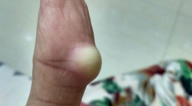
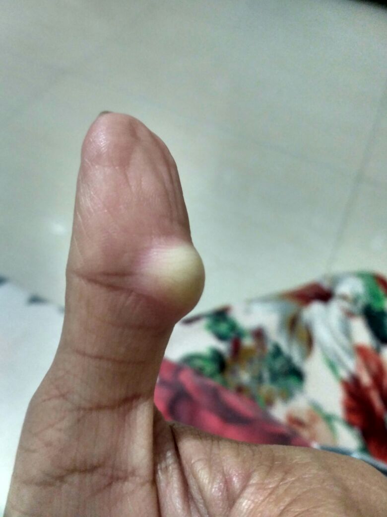
ONE OF OUR CASES OF GANGLION CYST CURED WITH HOMEOPATHIC MEDICINES WITHOUT ANY SURGERY
BEFORE HOMEOPATHIC TREATMENT
GANGLION CYST ON THUMB, BEFORE TREATMENT
AFTER HOMEOPATHIC TREATMENT
GANGLION CYST OF THUMB RESOLVED AFTER HOMEOPATHIC TREATMENT
SLE or Systemic Lupus Erythematosus or sometime only called Lupus is a chronic systemic autoimmune condition with genetics, nutrition and environment playing a major role in its onset and evolution, its a condition which greatly reduces life expectancy and also causes complications in pregnancy. It falls under disabling disease conditions category though much less than10% cases develops disabilities.
TYPES of SLE
Acute Cutaneous Lupus
Sub-Acute Cutaneous Lupus Erthematosus
Chronic Cutaneous Lupus also called Discoid Lupus Erthematosus
Neonatal Lupus Erythematosus
Drug Induced Lupus Erythematosus
EPIDEMIOLOGY of SLE
Lupus was the most google searched topic in healthcare related topics in 2017! Now that is something to be added in Epidemiology section! 😀 hehehe!
Female are affected more with SLE than male, female of child bearing age are affected more with female to male ratio of about 9:1.
African, Caribean and chinese ethnic groups are more prone to this condition.
SIGNS and SYMPTOMS of SLE
GENERAL
Undifferentiating symptoms which are common to other diseases as well.
Fever
Fatigue
Malaise
Joint pain
Muscle pain
Bodyache
Sleep disturbances
Psychiatric Disorders
Poor physical fittness
Anaemia of chronic disease
Raynaud’s Phenomenon
SKIN
Majority of cases shows skin manifestation of the condition
Though the rashes and lesion on skin may vary, the classic sign of SLE on skin is butterfly rashes on face, also called Mallar Rashes and is seen in almost half of the cases with skin lesions.
If it is Acute type there might not be scaling of skin, not well demarcated distinct edge compared to othe types.
If it is Subacute type rashes shows scaling of skin with distinct edges.
And in Chronic type rashes shows thick distinct thick scaling with very well demarcated edges.
Increased Hairfall is also one of the complaints.
Ulcers in mucous membranes esp of nose and mouth.
MUSCULOSKELETAL
It affects Small joints especially of fingers and wrist and it very closely mimicks rheumatoid arthritis and psoriatic arthritis making it clinically difficult in making differential diagnosis.
But it is less destructive and disabling compared to other two conditions, only less than 10% cases of SLE will develop deformities and even fewer will have disabilities.
It not only mimicks Rheumatoid Arthritis but it also seems to have very close relation to rheumatoid arthritis.
It significantly increases risks of fractures and also it is associated with facilitation of Osteoarticular Tuberculosis.
HEAMOTOLOGICAL
Anaemia of chronic disease with low Red Blood cell count
Low White Blood Cell count
Anti phospholipid antibody syndrome is a condition where anti-phospholipid antibodies increases Partial Thromboplastin time causing tendency to heamorrhages and it is frequently found positive in patient with SLE and its coexisting is termed as Lupus Anticoagulant Positive.
Patients with SLE frequently show positive results for Anti Cardiolipin Antibodies as well, and patients with these antibodies sometimes shows false positive results for Syphillis.
CIRCULATORY SYSTEM AND HEART
Artheroscelerosis – Thickening and deposition of cholestrol plaque in blood-vessel walls which may also give rise to Ischemic Myocardial diseases like Myocardial Infarction.
Vasculitis – Inflamation of blood-vessels in some cases
Endocarditis – Inflamation of inner linning of heart, when due to SLE its termed as Libman-Sacks Endocarditis
Pericarditis – inflamation of outer layers and and surrounding tissues.
Myocarditis – Inflamation of cardiac muscles.
It may also cause inflamation of Mitral and Tricuspid valves of heart.
LUNGS
Pleurisy – Inflamation of pleura.
Pneumonitis – Inflamation of lungs.
Interstitial Lung Disease
Pulmonary Embolism
Pulmonary Heamorrhage
All these complications and chronic inflamatory processes causes Shrinking Lung Syndrome where there is reduction in lung volume.
REPRODUCTIVE
30 % of pregnancy has comolications like
Fetal Death
Spontaneous Abortion
Still Birth
Prognosis is worse in those who get aggravations in SLE duringbpregnancy.
Neonatal Lupus Erythematosus
Child born to mother with SLE shows symptoms of Discoid Lupus Erythematosus with
Heart block
Splenomegaly – Enlargement of Spleen
Hepatomegaly – Enlargement of Liver
Neonatal SLE is self limiting condition and in most cases recovers on its own.
RENAL (Kidney)
Painless Heamaturia – Blood in urine.
Painless Proteinuria – Protein in urine.
Lupus Nephritis leading to terminal Renal failure.
Histologically it shows its classical appearance of Membranous Glomerulonephritis with Wire-Loop Abnormailities due to deposition of Immune Complexes in Basement Membrane.
NEUROPSYCHIATRIC (NP-SLE)
If any Neuro-Psychiatric is caused dur to SLE its called NPSLE Neuro-Psychiatric Systemic Lupus Erythematosus
There are atleast 12 Central Nervous System related and 7 Peripheral Nervous System relate manifestation of SLE that are being observed in patients with SLE.
SLE is considered amongst one of the prototype disease as its very difficult to differentiate from many other autoimmune conditions as they share in common majority of signs and symptoms making its diagnosis very difficult, It much depends on clinical picture of the disease and investigations after that there are many criterias based on clinical symptoms coupled with laboratory tests based on which a person can arrive at some conclusion though not absolutely sensitive and specific to confirm diagnosis in every case but fair enough.
LABORATORY TESTS
ANA detection by direct or indirect immunoflorescence
ANA test detects many different subtypes of ANA related to many other autoimmune conditions as well with many overlapping eachother of which
Anti- Double Strand DNA Antibodies most specific of all present in almost 70% cases of SLE with only 0.5% non-SLE cases has t in them.
Anti-Smith DNA Antibodies present in most of the cases of SLE and not frequently found in non-SLE person.
Anti- Histone Antibodies present in Drug Induced SLE
Anti- U1 RNP antibodies – non specific also appears in other conditions like Systemic Sclerosis
Anti- Ro or SS-A and Anti- La or SS-B – non specific for SLE but more Specific to Sjogrene syndrome, but its present in many of the cases of neonatal lupus with heart involvement in particular.
Other Tests
Anti-ENA Test
Lupus Cell Test – It was used in past as it used to show positive in 50-70% SLE cases but was not specific to SLE and used to be present in many cases of many other conditions like RA Scleroderma etc.
DIAGNOSTIC CRITERIA OF AMERICAN COLLEGE OF RHEUMATOLOGY.
Its a stringent criteria developed by American College of Rheumatology, so that non of non-SLE cases should filter in, so many of the SLE cases are also filtered out.
The criteria is that if any patient shows any of the four symptoms out of eleven simultaneously or serially in more than one occasion than he is considered to be positive for SLE.
Mallar Rash/ Butterfly rash on cheeks; Sensitivity of 57% and Specificity of 96%.
Discoid Rash; Sensitivity of 18% and Specificity of 99%.
Serositis, Inflamation of serous membranes around heart (more specific) and lungs(more sensitive); Sensitivity of 56% and Specificity of 86%.
Mucosal Ulcers of oral cavity and nasopharynx; sensitivity of 27% and specificity of 96%.
Arthritis, non-erosive with more than two joints involved with tenderness swelling and effusion; Sensitivity of 86% and Specificity of 37%.
Photosensitivity, Ligh causes aggravation in skin rashes or other Lupus related complaints; Senitivity of 43%and Specificity of 96%.
Non Drug Induce : Hemolytic Anaemia, Leucopenia, Lymphopenia, Thrombocytopenia; Sensitivity of 59% and Specificity of 89%.
More than 0.5g of total protein in urine in a day or cellular cast seen in urine under microscope; Sensitivity 51% and Specificity of 94%.
Anti- Nuclear Antibody positive; Sensitivity of 99% and specificity of 49%.
Positive Anti- Smith, Anti- Double Strand DNA, Positive Anti- Phospholipid Antibody, False Fositive Serological test for Syphillis; Sensitivity 85% and Specificity of 93%; Presence of Anti- ssDNA in 70% of cases.
Neurological disorder Seizure or psychosis; Sensitivity of 20% and Specificity of 98%
This is a very stringent criteria used for research purpose if we go through we may falsely conclude negative diagnosis and miss out on diagnosing many patients who are suffering from SLE
Aslo it misses out on certain factors like antiphospholipid anti bodies which has strong association with SLE there are many cases who are anti phospholipid antibody positive but are not fitting in above ACR criteria but still they are having SLE.
So more practicle approach widely used is through the Recursive Partitioning which has two classification trees
The Simplest Classification Tree – If patient has any immunological disorder with positive anti- Smith antibody, anti- DNA antibody, false positive serology test for Syphillis, presence of Lupus cells or Mallar rash/butterfly rash, then the person is diagnosed as positive for SLE; specificity of 92% and sensitivity of 92%.
Full Classification Tree : It uses six criterias; sensitivity of 97% and specificity of 95%
HOMOEOPATHIC MEDICINES FOR SLE
I have seen homoeopathy work wonders in SLE especially in cases with NPSLE because then the disease becomes very expressive about itself, it shows itself not only on skin and joints but also on Neuropsychiatric sphere which show various symptoms typically different in each individual and this is what is required in homeopathic medicine selection, that the body is expressing itself in mental sphere which makes remedy selection easier.
Always a proper case taking needs to be done in deep seated chronic autoimmune conditions like this and a deep acting polycrest remedy should be selected after proper repertorisation as per each individual constitution and constitutional treatment is the only permanant solution for such conditions.
Still some theraputic indications are given which can be helpful guide and can be used as per the symptomatology in course of disease if indicated intercurrently or during acute excerbations of disease showing following symptom.
BELLADONNA
– Typically suited in Mallar rash or Bitterfly rash of Syetemic Lupus Erythemotosua with symptoms of Neuro-Psychiatric SLE (NPSLE) where CNS involvement is markedly noted also suits in PNS symptoms of NPSLC
MERCURIOUS SOLUBILIS
Whenever in case of lupus there are oral and/or naso-pharyngeal ulceration this remedy is very well suited
BORAX
Again this is best suited in mucosal ulceration but in this remedy the ulceration are more marked in oral mucosa than in nasopharynx.
SYPHYLLINUM
A nosode a dose can be given intercurrent as anti miasmatic of the cases that shows syphillitic miasma in the background also useful in cases showing painless red mallar rash or butterfly rash with much thickening and exfoliation especially like in Chronic Lupus Erythemotosus, also suited well in ulcerations of oral and nasopharyngeal mucosa.
CINCHONNA OFFICINALIS
In cases with signs of hemolytic anaemia wether due to disease ot allopathic medicines, it will work wonders in both the cases.
FERRUM PHOSPHORICUM
Where the patient has febrile condition due to disease with malaise, fatigue, Hairloss and aneamia duw to lupus or its medicines, can also be givem in low potencies in biochemic form along with other medicines
FERRUM METALLICUM
Red acute rash typically in acute lupus erythematosus with involvement of oral mucosa, also in later chronic stages when there is are signs of anaemia of chronic disease of hemolytic anaemia
RHUS TOXICODENDRON
Works wonders in cases of lupus where it not only acts on skin but also wonderfully acts on the joints and musculoskelwtal complaints the disease shows.
HYOCYAMUS
In patients with symptoms of NPSLE.
ACONITE NAPELLUS
in acute violent spells of relaopse and aggravations with symptoms of NPSLE like mental restlessness and has fear fright and anxiety in general and fear of death in particular.