Category Archives: Dermatology

All posts related to dermatology and homeopathic treatment for dermatological complaints will be posted under this category.

ANAEMIA

Anaemia can be defined as decreased haemoglobin counts or reduced red blood cell counts or reduced oxygen carrying capacity of blood, due to “loss of” or “abnormality of” red blood cells or haemoglobin.

Normal Heamoglobin Counts

  • 6 months to 5 years of age > 11g/dl
  • 5 years to 12 years of age > 11.5g/dl
  • 12years to 16 years of age > 12g/dl
  • Adult Females (non-pregnant) > 12g/dl
  • Adult Females (pregnant) > 11gm/dl
  • Adult Males > 13g/dl

CAUSES OF ANAEMIA

  • Blood losss
  • Excessive Red Blood Cell destruction
  • Heamoglobinopathies
  • Hypovitaminosis B12
  • Hypoferremia
  • Anaemia of Chronic diseases
  • Autoimmune haemolytic anaemia
  • Inflamatory bowel diseases
  • Hypervolemia or water retention due to sodium or other salts.
  • Genetic hereditary conditions like Thalasemia
  • Certain cancers
  • Kidney diseases
  • Reduced erythropoetin production
  • Excessive RBC destruction
  • Impaired RBC production
  • Certain infections like malaria which causes RBC destruction.
  • Certain drugs which causes RBC destruction eg. Quinine causes chinchonism.
  • Bone Marrow lesions and pathologies
  • Etc.

CLASSIFICATION OF ANAEMIA

There are many types of anaemias. It can be broadly classified into 7 categories depending upon their causes

Anaemia due to

  1. Blood Loss
  2. Hemolysis
  3. Impaired or abnormal Erythropoesis
  4. Hypervolemia
  5. Chronic Diseases
  6. Nutritional deficiency

Based on RBC morphology it can be classified into 3 groups

  • Microcytic
  • Macrocytic
  • Normocytic

FEW COMMON and RARE TYPES OF ANAEMIA

  • Iron Deficiency Anaemia
  • Aplastic Anaemia
  • Megaloblastic Anaemia
  • Pernicious Anaemia
  • Sideroblastic Anaemia
  • Autoimmune Hemolytic Anaemia
  • Myelodysplastic Syndrome
  • Thalasemia
  • Fanconi Anaemia
  • Congenital Dyserythropoetic Anaemia
  • Daimond-Blacfan Anaemia
  • Myelopthisis
  • Anaemia of Prematurity
  • Erythroblastopenia or Pure Red Cell Aplasia
  • Hereditary Spherocytosis
  • Hereditary Elliptocytosis

SYMPTOMS

  • Weakness
  • Lethargy
  • In children it affects growth in general
  • Somnolence, Drowziness in day time
  • Disturbed sleep at night
  • Pallor, general pale appearance of skin, mucous membranes and eyes.
  • Dyspnoea on Exertion.
  • Reduced Immunity, tendency to catch infections and slow recovery and healing.
  • Bodyaches
  • Cyanosis in severe cases
  • Palpitations
  • Tachycardia
  • Low blood pressure
  • Chest pain
  • Depression
  • Craving for indigestible things , PICA
  • Cold clammy extremities
  • Oedematous swelling of extremities, dependent oedema
  • Angina or cardiac failure in severe cases
  • Will impact general growth and repair of all the vital organs and tissue of the body.

HOMEOPATHIC MEDICINES FOR ANAEMIA

Depending upon the cause of anaemia and general constitution of the patient, one of the following medicines may be called for duty by a homeopathic physician.

  • Ferrum Metallicum
  • Ferrum Phosphoricum
  • Cinchonna Officinalis
  • Natrum Muriatic um
  • Arsenicum Album
  • Abrotanum
  • Hamamelis Verginiana
  • Pulsatilla Nigricans
  • Janosia Ashoka
  • Crotalus Horridus
  • Lachesis
  • Acidum Phosphoricum

MYCOSIS FUNGOIDES

Mycosis Fungoides is a type of Non-Hogkin Lymphoma. It is the most common type of Cutaneous T-Cell Lymphoma(CTCL).

Mycosis Fungoides is a misnomer which means “Mushroom-like fungal disease” but by no means it is a fungal condition. It is a type of blood cancer caused due to unusual expression of Skin-associated CD4 T-Cells. This cancer affects skin and produces various lesions on skin and rarely metastatise in other tissues.

It is a fatal condition with 10year survival rate of less than 70% which is even less in elderly.

It usually appears in people above 20years of age and is more common after age of 50yrs , males are affected more than females, the disease tends to be more fatal in black race. Survival rate has been found good in married white women.

CAUSES

Causes of Mycosis Fungoides remains unclear

No hereditary or genetic factors have been found associated to the disease.

It is considered to be non-contagious but some studies suggest that Human T-cell Lymphotropic Virus is associated with this condition.

SYMPTOMS

Initially disease shows symptoms that are not distinguishing and in most of the cases it resembles psoriasis or eczema. Even on biopsy it can not be diagnosed easily in early stages. Even in later stages multiple biopies are required to establish diagnosis.

Disease presentation begins from skin as exfoliating, pruritic, erythodermic eruptions resembling to Eczema or Psoriasis and is almost always mistaken initially in most cases. These eruptions tends to appear first on buttocks in vast majority of cases and then spreads throughout the body. Pruritus is present in only 20% of all the cases. In later stages tumourous and ulcerative lesions may also appear. Disease usually in most cases starts appearing at around age of 45-50 and is a slow prohressing condition with and its usually around age of 60+ that mist patient starts presenting symptoms of later stages like tumours, generalised pruritus and erythrodrema and ulceration. Though in many cases it may start as early as age of 20 and rarely before 20yrs of age. The diseases is classified into three stages

Stages of Mycosis Fungoides

Mycosis fungoides is divided into three stages viz.

  1. Pre-Mycotic
  2. Mycotic
  3. Tumourous

Pre Mycotic Stage

It starts appearing on skin at this stage as erythematous, pruritic, exfoliating skin eruptions.

On biopsy it cannot be diagnosed at this stage as histopsthology shows pattern of non specific dermatosis with epidermal psoriasiform changes.

Mycotic Stage

In Mycotic stage Infiltrative Plaques starts appearing as slightly raises or wrinkled spots.

On histopathology polymorphs and few atypical lymphoid cells are seen inflamatory infiltrates in dermis. When this cells are seen lined up.in epidermal basal layer without spongiosis then its confirmatory finding to establish diagnosis of mycosis fungoides.

Tumorous Stage

Nodular overgrowths are seen on skin ranging from few milimeters to centimeters scattered throughout the body along with plaques and patches. Usually it appears on the same place where the plaques and patches are. Also there is erosion of the tumours and ulcerations is seen in many cases over the tumour and plaques.

On histopathology they show medium sized lymphocytes with cerebroid nuclei expands dermis.

DIAGNOSIS OF MYCOSIS FUNGOIDES

Diagnosis is based primarily on histopathological findings of the skin lesions

Clinically patients showing features suggestive of or doubted mycosis fungoides are closely monitored over the time untill biopsy confirms. Multiple boipsies of various lesions might be required at every progressing stage untill confirmed, as its very difficult to estsblish diagnosis in initial stafe wether clinically or histopathologically as it resembles psoriasis or eczema.

On histopathology following findings are required to establish diagnosis of mycosis fungoides

  • Band-like lymphocyte infiltrate in superficial papillary dermis
  • Epidermotropism
    • Cerebroid T-cells in dermal and epidermal infiltrate
  • Pautriers Microabscesses – though not present in most cases but is characteristic finding of mycosis fungoides where in the epidermal infiltrate shows atypical lymphocytes arranged in aggregates of four or more.

HOMOEOPATHIC MEDICINES FOR MYCOSIS FUNGOIDES

  • Thuja Occidentalis
  • Syphillinum
  • Antimonium Crudum
  • Arsenicum Album
  • Ars Sulph Flavum
  • Phosphorus
  • Argentum Nitricum
  • Hepar Sulphuris
  • Sulphur
  • Silicea

TINEA INFECTIONS DERMATOPHYTOSIS

Tinea is the term used to describe a group of contagious skin infections caused by different type of fungi.

Dermatophytes are a group of organisms that are able to breakdown the skin tissues.

Dermatophytosis is mainly caused by microsporum, trichophyton and epidermophyton.

These dermatophytes grow best in warm and humid environments.

DIFFERENT TYPES OF TINEA INFECTIONS

It affects many areas of skin and depending on thier location and fungal infection it has different names.

Common type of Dermatophytosis depending on the sites are as follows
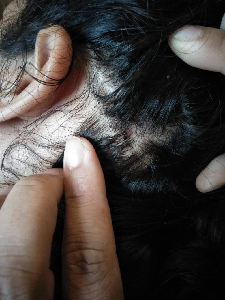
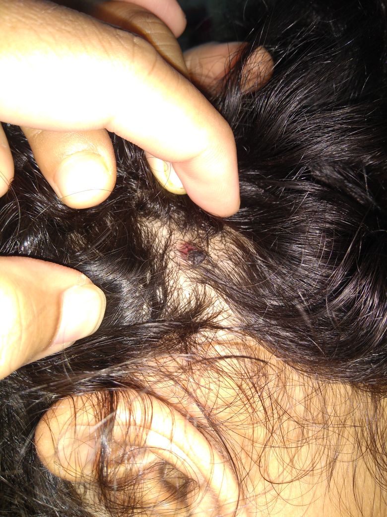
TINEA CAPITIS

Scalp ringworm causes itchy patches on the head, it can leave bald spots, usually affects children.

TINEA BARBAE

It is the infection of the skin especially beard area and moustache and is usually seen in men.

TINEA CRURIS

It is also known as Jock’s itch. Infection of groin, vulva, inner thighs , buttocks.
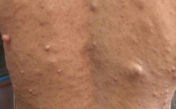
TINEA CORPORIS

When Dermatophystosis presents on the parts of body like trunks, hands, feet etc.

TINEA UNGUIUM

It is infection of one or more finger nails or toe nails.

TINEA PEDIS

It is also known as Athlete’s foot, causes infection of the foot or inter digits of foot.

CAUSES OF TINEA INFECTIONS

Dermatophytosis are the group of organisms that are able to break down the skin tissue. The fungi reside in the soil and are involved in decomposition and can infect the living organisms.

Dermatophytes are transmitted from person to person and some are adapted to animals.

Dermatophytosis is mainly caused by microsporum, trichophyton and epidermophyton. These dermatophytes grow best in warm and humid environments.

It can be easily transmitted through skin to skin contact with an infected person or animal or through indirect contact with an object or surface that an infected person or animal has touched.

Bathroom floors, bathmaths, towels, showers and communal bathing, swimming and changing room areas are common source of infection.

On contact with skin, the dermatophyte spread to the surface layers of the skin.

SYMPTOMS OF TINEA INFECTION

Symptoms of the fungal infections vary depending upon the infection and affected tissues and area of the body.

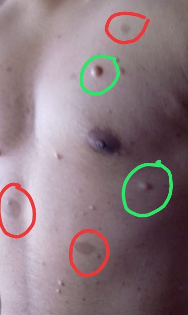
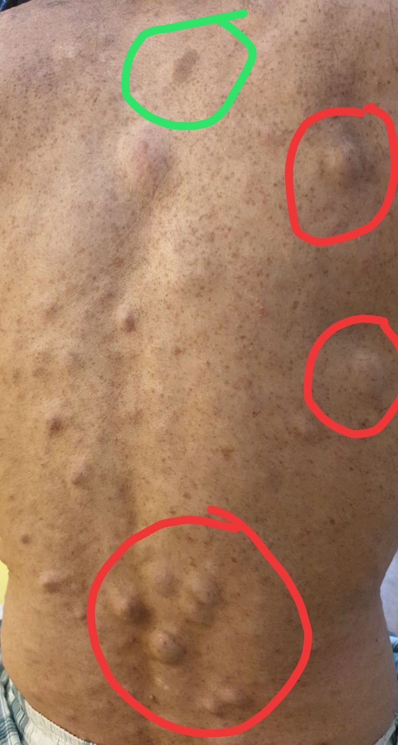
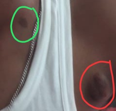
The skin lesions are usually characterised by redness, inflammation that is more severe at the edges with raised and thick edges, it is usually seen that the central area is clear.

SYMPTOMATIC REPRESENTATION OF TINEA DEPENDING ON THE AFFECTED SITE
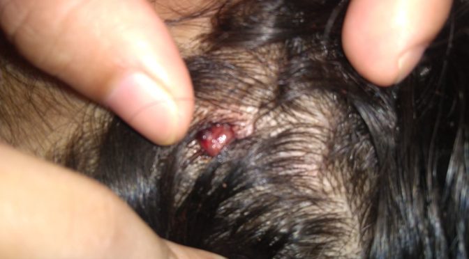
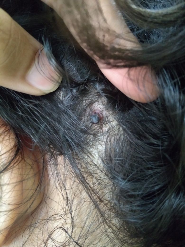
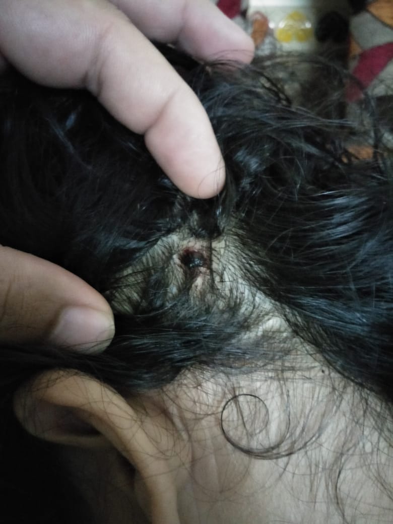
TINEA CAPITIS

Tinea Capitis is also known as scalp ringworm. It usually affects children. Seen in children with infection of hair and scalp. The infection is characterised by scaly, irregular or well marked redness with patchy hair loss, the patchy areas are dry with loss of hair and minimal inflammation.

TINEA BARBAE

The dermatophytic infection that involves hair and skin of the face that is the beard and moustache. The lesions may include scaling with pus along with redness.
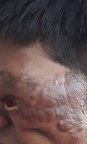
TINEA FACIEI

It is seen on non beard parts of the face.

TINEA CRURIS

This is an acute to chronic infection of the groin and adjacent areas like inner thighs, butocks. The symptoms usually imclude burning, severe itching and redness with scales, raised edges, sharply demarcated borders with clear centre. Skin becomes dark in the centre as the lesion progreses.

TINEA CORPORIS

Tinea Corporis occurs on the trunk, hands and feet. The infection usually tends to spread to other areas. The lesions are usually pink to red or scaly and annular with slightly raised and sharp margins with clear centre. Sometimes lesions may present with pus.

TINEA UNGUIUM

Tinea Unguium is infection of nails characterised by thickened opaque, a discoloured or deformed nails. The nail may be separated from nail bed. Toe nails are more affected than finger nails.

TINEA PEDIS

Tinea Pedis is seen between the digits of feet. Commonly called as athlete’s foot. There is dryness, fissures and scales, moist lesions in some or all spaces. The lesions may be present with scaling of the soles with inflammation and dryness or redness.

ROLE OF HOMEOPATHY IN TINEA INFECTIONS

Homeopathic treatment is very beneficial and effective mode of treatment that boosts the bodys immune process to fight and eradicate fungal infections.
As the infection gets older, it tends to get more resistant to any form of treatment, once external applications are used for months together the infection becomes resistant. Homeopathy plays an important role in such cases.

INDICATED HOMEOPATHIC REMEDIES

SULPHUR

Sulphur is very beneficial homeopathic medicine for various skin diseases. In cases of ring worm lesions sulphur is quiet useful.
The patient presents with intense itching and burning in erruptions. Indicated in cases of infections that have been suppressed by local medications of various kinds.

PSORINUM

Psorinum is one of the indicated remedy for skin infections. Mainly used for ringworm infections of scalp and bends of joints. There is severe itching in the affected area. Itching worse due to warmth of bed. Indicated in tinea capitis. The hair appears dry, rough, and lustreless. Profuse sweating with offensive body odour throughout the body.

SEPIA

Indicated in case of fungal infections appearing in isolated parts of body. Infections can occur on any part of the body, but common location is bends of knees and elbow. The erruptions are accompanied by itching and scratching, no relief from scratching. High senditivity towards cold air.
Irritable, indifferent towards family and friends.

THUJA

Thuja is indicated in skin infections. Main action is on skin. Indicated in case of dermatophytic infection where the erruptions are on the covered parts of the body, worse from scratching, very sensitive to touch. Worse at night from heat of bed, from cold damp air. Scratching and cold bathing makes the condition worse.

GRAPHITES

Graphitis is one of the indicated remedy for jocks itch Tinea Cruris with recurrent episodes. Indicated where the skin is dry, rough with severe itching. Sometimes moist crusty erruptions are seen on the parts. There is oozing of sticky exudation. Graphitis is effective for fungal infection around vulva.

ARSENIC ALBUM

Arsenic album is beneficial in Tinea Capitis. The scalp shows bald spots with intolerable itching with intense burning. The symptoms are usually worse at night. Arsenic alb helps to reduce itching and burning and also helps in regrowth of hair on bald spots.

SILICEA

Silicea is indicated in Fungal infections and dermatophytosis in Delicate pale waxy patient. Scars are painful. Eruptions itch only in the day time and evening. Crippled nails. Indicated in case of Tinea Pedis with offensive sweat, deep cracks between toes with intense itching and burning. Painful foul smelling ulcers of the foot.

PERTROLEUM

Petroleum is indicated in case of Tinea Pedis. Helps to drive away infection of foot. Presents with offensive sweat. Deep cracks between toes with intense itching and burning of skin. Crippled and brittle nails.

TELLURIUM

Tellurium is indicated in Dermatophytosis with lesions on large parts of the body. Usually it covers a broader area of skin. Presents with excessive itching and stinging sensation. Itching day and night. Cold air worsens the itching. Offensive odour from the affected area.

CHALAZION

WHAT IS CHALAZION?

Chalazion is a small, usually painless lump or swelling that appears on eyelids as a result of cyst formation due to blockage of duct of Meibomian gland or rarely of Glands of Zeis. It can appear on upper and lower eyelids. It can be single or multiple and can affect both the eyelids of both the eyes at same time.

CAUSES AND RISK FACTOR

CHALAZION is caused by a blockage in duct of one of the tiny Meibomian Gland of upper and lower eyelid or due to infection or inflammation of Meibomian Gland and rarely of Gland of Zeis in which it is usually secondary to Stye.

Meibomian Glands are responsible for secretion on Meibum which is oily in nature that traps tear within eye by forming thin layer above surface of secreted tear, thus retaining it in eye by not letting it spill out and also reduces evaporation of tears thus playing a major role in keeping eye moist. There are around 50 mebomian glands in upper eyelids and 25 mebomian glands in kower eyelids they are situated on the rim of eyelids on Tarsal plate

Glands of Zeis are sebaceous glands that open up in hair follicle of eyelash they secrete sebum to keep eyelids and eyelash soft and supple, it also plays part in providing lubrication. In this gland the chalazion formed is usually secondary to stye and is not too frequent.

It is more common in people with following risk factors

  • Chronic Blepharitis
  • Seborrhea
  • Rosacea
  • Long term inflammation of eyelids.
  • Infective conjunctivitis.
  • Person wearing contact lenses.
  • Those who have frequent exposure to pollution, smoke, dust etc.
  • Those using Kajal or Surma.

SYMPTOM OF CHALAZION

  • Painless lump or swelling in upper and lower eyelid usually visible or palpable.
  • It can affect both eyes at the same time.
  • Usually they are well defined round with sharp margin and whole surface of sphere is easily palpable if held netween fingers with eyelids gliding over it.
  • There can be one or multiple chalazia.
  • Sometimes in cases of multiple the chalazia, due to frequent secondary infections, they may mate and overlap adjecent chalazia.
  • In mated and overlaping multiple chalazia it becomes necessary to rule out malignancy.
  • Symptoms depend on its size larger chalazia may cause disturbance in vision due to obstruction of viewing fleid.
  • In some cases it may cause nystagmus.
  • If there is secondary infection then it  might be red and inflammed in appearance and may also occasionally be painful.
  • May complicate and show blepheritis as well.

DIAGNOSIS

Sometimes Chalazion may be confused with external or internal Stye.

DIFFERENCE BETWEEN STYE AND CHALAZION.

EXTERNAL STYE

Is caused due to infection in the area of eyelash follicle or sweat gland.

INTERNAL STYE

It is caused due to infection of a meibomian gland.

Difference between chalazia and stye is that stye are painful but chalazia usually aren’t and chalazia may develop after stye.

By physical examination or by asking the patient if there is pain so that it can be differentiated whether it is stye or chalazion. Although chalazion with secondary infection may be painful, so proper evaluation is required in all doubted cases, by checking the location and palaption helps to diagnose properly.

Chalazia those are abnormally large or has abnormal texture of skin over it or is fixed and skin over it is not freely gliding or they are recurring or multiple or overlaping or suspicious with other unusual characteristics should be investigated for malignancy.

Differential diagnosis includes

  • Stye
  • Mebomian adenoma
  • Carcinoma of sebaceous gland
  • Sarcoid Granuloma
  • Foreign Body Granuloma.

TREATMENT

Chalazion may disappear by its own just use a warm compression to your eyelid it can reduce the swelling. If it does not reduce then necesary treatment is essential.

ROLE OF HOMEOPATHY:

Under conventional mode of treatment, surgical removal of chalazion is the only option. which has its own comolications like dryness of eyes and irritation of eyes due to scar formation at site of excision and also it damages and makes mebomein glands non functional forever.

However, In homeopathy chalazion is treated in a non invasive manner with gentle and effective approach.

Few indicated homeopathic remedies in Chalazion are

APIS MELLFICA

Presents with swollen eyelids, redness, intolerance of heat and touch especially right sided with burning and stinging sensation.

PULSATILLA NIGRICANS

Patient presents with itching and thick profuse discharge, Worse in warm room better in open air. There is dimness of vision swelling of eyelid and patient wishes to rub or wipe eye constantly.

STAPHYSAGRIA

Indicated in Recurrent chalazion, better by warmth application. Sunken eyes , heat in eyeball, feel dry despite lachrymation.

THUJA OCCIDENTALIS

Indicated in case of Chalazion. Eyelids stick together, usually left sided, eye lid feel heavy as lead and may be dry and scaly. Bluring of vision.

GRAPHITES

Indicated in case of chalazion, recurrent stye, conjunctivitis, red and swollen eyelid with much dryness it is good for blepharitis too. Indicated remedy for eruption on lids that become hard over a period of time.

CALCAREA FLOURICA

Indicated in case of chalazion. Gland enlarged and become stony hard useful in patient has had chalazion for a very long time. Swelling of eyeild with pain.

CONIUM MACULATUM

Indicated in case of chalazion. Frequently used remedy for stye and chalazion which have a tendency to become stony hard over a period of time for chronic and long standing complaints.

HEPAR SULPHURIS

I have found this remedy effective in almost allthe cases of chalazion when given in 30 potency with frequent repetition 2-3 times a day for a prolonged period of time and then chalazion starts showing signs of inflamation after which the medicines should be stopped and soon we see the chalazion gradually is resoved in few weeks after the medicines were stopped.

PREMATURE GREYING OF HAIR

There was a time when premature greying of hair happend to only older people. Premature greying of hair has become a cause of concern for younger generation. Its not uncommon anymore to see people in thier early 20’s with a mop of grey hair on thier head. Premature greying of hair has become an important concern in present day and thousands of people both men and women alike are seeking medication for the same. Premature greying of hair can have a negative effect on appearance, confidence and social acceptance of the affected individual.

HOW AND WHY EXACTLY DOES GREYING OF HAIR OCCURS?

The root of every hair strand under the scalp is surrounded by a hair tissue called hair follicle which contains pigment cells. These cells produce the chemical called melanin that gives hair strand its colour.

When a person grows older the melanin production reduces making the strand greyish or white.

WHAT CAUSES PRE MATURE GREYING OF HAIR?

It is usually a complex multifactoral process.

Genetic factor: is one of the most common reasons for having grey hair at an early age.

Stress: Person under prolonged stress anxiety tension can be one of the cause of greying at an early age.

Dietary deficiencies: Deficiency of vitaminB, zinc, copper. These nutrients assist the metabolic process involved
in giving hair its natural colour.

Smoking damages the melanin producing cells melanocytes causing greying of hair.

Thyroid disorder: An underactive or overactive thyroid can affect the melanin production of hair.

HOMEOPATHIC APPROACH IN PREMATURE GREYING OF HAIRS

It is important to find the underlying cause for greying of hair.
Once this is done,the condition is best attended by Homeopathy.
With effective counselling,diet and regimen followed by a homeopathic medicine really helps.

HOMEOPATHIC REMEDIES INDICATED IN PREMATURE GREYING OF HAIRS

PHOSPORIC ACID
LYCOPODIUM
NATRUM MUR
PHOSPHORUS
SILICEA
VINCA MINOR

PREMATURE GREYING OF HAIR THERAPEUTICS :

EMBELICA OFFICINALIS or AMLAKI

Since ages Amla is known to benefit health of hair. Amla is rich source of Vitamin C, Iron, Calcium and has lots Fibres. In ayurveda it is well proven that amlaki helps reduce greying of hair Similarly in Homeopathy the tincture prepared from amla that is Embelica Officinalis Q also called Amlaki Q is proven to be too beneficial in cases of premature greying of hairs.

PHOSPHORIC ACID

Phosphoric acid is one of the indicated remedies for the treatment of premature greying of hair resulting from grief and depression. There is thining and falling out of hair. The patient is indifferent to all affairs of life.Phosphoric acid helps to restore the pigmentation of hair that are destroyed by effects of grief.

NATRUM MURIATICUM

Natrum muriaticum is indicated in treatment of premature greying of hair mainly in weak and anaemic person. Natrum Mur helps to improve general health of the patient and raise immunity to combat greying of hair. Natrum patients have severe headache which gets worse in the heat of sun.

SILICEA

Silicea is very useful in treating grey hair when the digestive power and absorption of nutrients is weak. Deficiency of nutrients lead to greying of hair. Silicea helps to improve the proper absorption of nutrients and hence prevent greying of hair.

PHOSPHOROUS

Phosphorous is indicated in treatment of grey hair. It prevents further greying of hair. The patient presents with hairfall and dandruff, itchy dry scalp. Desires icy cold drinks.

VINCA MINOR

Indicated in case of greying of hair. Helps to prevent greying of hair. Indicated when hair falls in spots, vinca helps to restore natural pigment of hair.

LYCOPODIUM

Indicated in case of premature greying of hair. Lycopodium halts the process of greying of hair when de pigmentation of hair occurs in spots. Indicated in patients whose hair problems are associated with gastric disturbances.

THYROIDINUM

Indicated in premature greying of hair along with thyroid disorders. Helps in arresting the process of greying of hair.

CALCAREA CARBONICA

Indicated in greying of hair presents with severe hair loss, sweating of scalp followed by intense itching.

DIETARY ADVICE FOR CASES OF PREMATURE GREYING OF HAIR

  • Increase your intake of fruits rich in vitamin C.
  • Amla since ages is known to aid in cases of premature greying of hair, it is recomended to consume atleast one amla everyday and when off season on can purchase dried amla powder. Amla Hair oil for external application on hair and scalp is also known to prevent greying of hairs.
  • Make sure your diet contains sufficient amount of green leafy vegetables
  • Adding almonds and other nuts rich in vitamin E can help reduce greying of hair.
  • Walnuts contain biotin that helps strengthen hair, reduces occurance of premature grey hair.

PYOGENIC GRANULOMA

What is Pyogenic Granuloma?

PYOGENIC GRANULOMA also called GRANULOMA TELANGIECTATICUM or LOBULAR CAPILLARY HEAMANGIOMA is relatively a common benign vascular lesion of skin and mucosa and appears as an overgrowth of tissue.

It is usually a small, round, bloody red in color.

They are also known as Lobular capillary haemangioma or granuloma telangiectaticum.

They grow rapidly at first and then remain a constant size, they rarely exceed 1cm in diameter.

What causes Pyogenic Granuloma?

The following factors have a possible role in their development

Some cases develop at the site of a recent minor injury, such as a pin prick.

Staphylococcus aureus is frequently present in the lesion.

Hormonal changes in the body during pregnancy can also be the cause of Pyogenic Granuloma.

Certain drugs and medications are known to cause pyogenic granuloma

SIGNS AND SYMPTOMS

The appearance of Pyogenic Granuloma is usually a small red, round, oozing and bleeding bump.

Younger lesions bleed easily because they are rich in blood vessels. These lesions can be painful.

The lesions arise on various regions of the body such as eyelids, face, tongue, lips, arms, hands and feet.

EYELID: The condition gives rise to reddish pink vascular bumps over the eyelid and is said to be caused due to a rich blood supply from conjunctiva.

TONGUE: These growths arise on lateral side of the tongue, it may arise due to trauma from dentures.

LIP: Arises as a solitary growth on the lip.Growth generally appears on the lower lip.

FINGERS:The growths are common on hands and fingers.

FACE: Growths arising on face are generally multiple in number and looks like small red or reddish purple lumps

FEET: These bumps arise in the nail bed of a toe with an accompanying ingrown toe nail.

DIAGNOSIS

Pyogenic Granuloma can be diagnosed based on its appearance.

HOMEOPATHIC MANAGEMENT

Commonly Used Homeopathic Medicines for Pyogenic Granuloma

SYPHILLINUM

Syphillinum is mostly indicated in case of pyogenic granuloma, where the patient presents with abnormal growths with pain. Indicated in case of chronic erruptions. Suited to person with pale and fine textured skin, who are slender. Presents with marked prostration and debility.

HEPAR SULPH

Hepar sulph is indicated in pyogenic granuloma. Specially suited to lymphatic and phlegmatic individuals. Excessive sensitiveness of parts is a leading indication. Chilly sensation. Indicated in csae of vascular lesions of the skin, they are bright red in colour which tend to bleed.

SILICEA

Silicea is mainly indicated in cases like pyogenic granulomas. Patient presents with vascular lesions of the skin where the suppurations continue and wound refuse to heal. Discharge is thin bloody. The lesion are benign and granulomatous.

THUJA OCCIDENTALIS

Thuja is indicated in case pyogenic granuloma as it is used in bleeding growths, spongy tumours, warts. Thuja has main action on the skin. The lesions have tendency to bleed easily and presents with pain. Complaints worse at night.very sensitive to touch.

RUTA GRAVEOLENS

Ruta is indicated in case of pyogenic granuloma. The patient presents with small rounded vascular lesions which tend to bleed easily. Very anxious and low spirited with mental dejection. Presents with neuralgia, burning stinging pain.

CALCAREA FLOURICA

Indicated in case of pyogenic granuloma. Vascular lesions which tend to bleed and are painful. Indicated in abnormal grwoths. There is great depressio . Sensitiveness to cold. Mostly indicated in blood tumors.

Homeopathic Approach in case of Pyogenic Granuloma

In Homeopthy we not only consider the signs and symptoms of the patient but treat the patient holistically, based on individualisation.

Similarly considering the symptoms-similarity, individuality and total of the patient, below is an example of one of our cases of pyogenic granuloma at Dr SHAH’s HOMOEOPATHY this case responded well to homeopathic medicine Syphillinum and got completely resolved with selected potency of 200C of the said remedy.

pyogenic granuloma
Pyogenic Granuloma Before treatment

Pyogenic Granuloma
During Homeopathic treatment Pyogenic Granuloma becoming dark and dry

PYOGENIC GRANULOMA
DURING HOMEOPATHIC TREATMENT  PYOGENIC GRANULOMA BECOMING DARKER DRIER AND SHRINKING IN SIZE

DURING TREATMENT – SEE THE LARGE FRAGMENT OF PYOGENIC GRANULOMA HAS GOT DETACHED AND HAS INTERMINGLED IN HAIR NEAR INDEX FINGER

Pyogenic Granuloma
During Homeopathic Treatment at Dr Shah’s Homeopathy

Pyogenic Granuloma resolved after detaching and shrinking completely now only a small dark spot can be seen at the site.

Pyogenic Granuloma
Pyogenic Granuloma- After Treatment – Cured completely after Homeopathic Treatment By Dr DEEPAN P SHAH at Dr Shah’s Homoeopathy.

ORAL MUCOCELE cured with Homeopathy

What is Oral Mucocele?

Oral Mucocele is a cyst formed due to collection of salivary gland secretion.

It is pearly translucent occasionally with bluish discoloration usually measuring from 1-8mm in diameter. The deeper cysts may appear as a nodule and may not have translucent pearly appearance as in superficial cyst.

It appears on lower lip on sides.

When it appears on floor of mouth below tongue its termed as ranulla.

They are painless most of the time.

It usually lasts for few days to years.

It may frequently rupture and reappear again at same site.

It may also cause ulcerations.

Oral Mucocele
Oral Mucous retention cyst

Oral Mucocele, Mucous Retention Cyst, Mucous Extravasation Phemenon

Oral Mucocele results from

  • Ruptured Salivary Gland Duct this results into mucous extravasation phenomenon.
  • Obstructed Salivary Gland Duct this results into mucous retention cyst.

Rupture or Obstruction can be due to

  • Infection that can potentially obstruct or damage the salivary gland and its duct.  which can be due to many factors most common being poor oral and dental hygiene.
  • Injury on lower lip which can damage salivary gland duct may result innto oral mucocele tje most common form of injury is due to bitting lips by one own teeth.
  • Tumours that may obliterate obstruct or rupture or damage salivary glands and its ducts in any form may result in to mucocele in oral mucosa

HOMEOPATHIC TREATMENT FOR ORAL MUCOCELE

The underlying cause needs to be evaluated, exclude the probability of infection, malignancy. Patient needs to be guided for proper oral hyegiene, any habbit like bitting teeth should be avoided.

Following are the common Homeopathic  medicines used in treatment of Oral Mucocele

  • Silicea
  • Thuja
  • Variolinum
  • Calcarea flourica
  • Mercurious Solubilis
  • Syphyllinum
  • Antimonium Tartaricum
  • Antimonium Crudum
  • Apis Mellifica
  • Ranunculus bulbosus

Selection from the above medicines should be strictly done as per symptom similarity basis under consultation of homeopathic practitioner.

NEUROFIBROMATOSIS (NF)

NEUROFIBROMATOSIS (NF)

Neurofibromatosis is syndrome of Phakomatosis or Neuro-Occulo-Cutaneous type of Autosomal Dominant Genetic Anomaly. It can be hereditary germline change or acquired during early developmental stage, affecting structures of embryonic ectoderm, where in, there is mutation or rarely somatic mosaicsism(RASopathy) of NF1 gene or its alleles, present on Chromosome 17 and is responsible for synthesis of protein neurofibromin which regulates cell proloferation and differentiation which hampers tumour suppressor activity giving rise to tumours of nervous system.

TYPES OF NEUROFIBROMATOSIS

  1. NEUROFIBROMATOSIS TYPE 1. 

    It is considered to be syndrome falling under category RASopathies. It affects supporting structures of Peripheral Nervous System. This type rarely show any severe complication other than cosmetic and compression complaints.

  2. NEUROFIBROMATOSIS TYPE 2.  

    Involves Schwaan cell of Cranial nerves especially of Vestibulocochlear nerve that is 8th cranial nerve or CN VIII causing bilateral acoustic Schwannoma causing hearing loss. It shows many complications of cranial nerves and brain stem. It can be disabling and life threatening. Severeity varies in different individuals depending on the way in which the disease assumes its course in different individual.

  3. SCHWANNOMATOSIS.   

    In this type Schwann cell tumours are found in Peripheral and Spinal nerves. It can be very painful and disabling.

SYMPTOMS OF NEUROFIBROMATOSIS

  • Multiple upto six or more “cafe au lait” spots more than 5mm in diameter is one of the diagnostic sign which are also present in Mc Cune Albright Syndrome.
  • Freckles generalised dispersed throughout body increasing simultaneously with neurofibromas.
  • Excessive folds of skin especially in underarm region with freckles.
  • More than one overgrowths in iris of eye.
  • More than one Neurofibroma.
  • Spinal growth and curvature anomalies.
  • During early stages of development in children it may show Learning and Behavioral problems

Cafe au Lait spots and neurofibroma in patient with Neurofibromatosis. 1)Neurofibroma marked in Green Circles 2)Cafe au Lait spots marked in Red Circles
Cafe au Lait spots and neurofibroma in patient with NF
1)Neurofibroma marked in Green Circles
2)Cafe au Lait spots marked in Red Circles

Neurofibromatosis patient with classical presentation of Cafe au Lait Spot, Neurofibromas and extensive freckles on back.
1)Café au Lait spot – dark brown spot marked in green circle
2) Neurofibroma – nodules marked in red circles
3)Freckles – Extensively scattered, small dark dots like pigmentation throughout the back
Above three are common findings in patients with Neurofibromatosis.

Plexiform Neurofibroma on eyelids in patient with neurofibromatosis
Plexiform Neurofibroma of eyelids

Cafe au Lait spots and neurofibroma in patient with Neurofibromatosis. 1)Neurofibroma in Red Circle 2) Cafe au Lait Spot in Green circle
Cafe au Lait spots and neurofibroma in case of NF.
1)Neurofibroma in Red Circle
2)Cafe au Lait Spot in Green circle

DIAGNOSIS OF NEUROFIBROMATOSIS

  • HISTOLOGY helps to confirm neurofibroma
  • SLIT-LAMP TEST
  • GENETIC TESTING helps confirm the diagnosis.
  • EEG, MRI, CT SCAN AND MRI helps us to assess tumour and its influence on surrounding tissue
  • Sphenoidal dysplasia on imaging, is one of the major finding in diagnosis  of NS1

HOMEOPATHIC MEDICINES FOR NEUROFIBROMATOSIS

  • THUJA
  • HYPERICUM
  • SILICEA
  • MEDHORRINUM
  • CONIUM MACULATUM
  • COCCULUS
  • BELLADONNA
  • CALCAREA FLOURICA
  • CALCAREA CARBONICA
  • GELSEMIUM SEMPERVIRENS
  • RUTA GRAVEOLENS
  • SYMPHYTUM
  • CALCAREA PHOSHORICA

SLE SYSTEMIC LUPUS ERYTHEMATOSUS

SLE or Systemic Lupus Erythematosus or sometime only called Lupus is a chronic systemic autoimmune condition with genetics, nutrition and environment playing a major role in its onset and evolution, its a condition which greatly reduces life expectancy and also causes complications in pregnancy. It falls under disabling disease conditions category though much less than10% cases develops disabilities.

TYPES of SLE

  • Acute Cutaneous Lupus
  • Sub-Acute Cutaneous Lupus Erthematosus
  • Chronic Cutaneous Lupus also called Discoid Lupus Erthematosus
  • Neonatal Lupus Erythematosus
  • Drug Induced Lupus Erythematosus

EPIDEMIOLOGY of SLE

Lupus was the most google searched topic in healthcare related topics in 2017! Now that is something to be added in Epidemiology section!  😀 hehehe!

Female are affected more with SLE than male, female of child bearing age are affected more with female to male ratio of about 9:1.

African, Caribean and chinese ethnic groups are more prone to this condition.

SIGNS and SYMPTOMS of SLE

GENERAL

Undifferentiating symptoms which are common to other diseases as well.

  • Fever
  • Fatigue
  • Malaise
  • Joint pain
  • Muscle pain
  • Bodyache
  • Sleep disturbances
  • Psychiatric Disorders
  • Poor physical fittness
  • Anaemia of chronic disease
  • Raynaud’s Phenomenon

SKIN

Majority of cases shows skin manifestation of the condition

  • Though the rashes and lesion on skin may vary, the classic sign of SLE on skin is butterfly rashes on face, also called Mallar Rashes and is seen in almost half of the cases with skin lesions.
  • If it is Acute type there might not be scaling of skin, not well demarcated distinct edge compared to othe types.
  • If it is Subacute type rashes shows scaling of skin with distinct edges.
  • And in Chronic type rashes shows thick distinct thick scaling with very well demarcated edges.
  • Increased Hairfall is also one of the complaints.
  • Ulcers in mucous membranes esp of nose and mouth.

MUSCULOSKELETAL

It affects Small joints especially of fingers and wrist and it very closely mimicks rheumatoid arthritis and psoriatic arthritis making it clinically difficult in making differential diagnosis.

But it is less destructive and disabling compared to other two conditions, only less than 10% cases of SLE will develop deformities and even fewer will have disabilities.

It not only mimicks Rheumatoid Arthritis but it also seems to have very close relation to rheumatoid arthritis.

It significantly increases risks of fractures and also it is associated with facilitation of Osteoarticular Tuberculosis.

HEAMOTOLOGICAL

  • Anaemia of chronic disease with low Red Blood cell count
  • Low White Blood Cell count
  • Anti phospholipid antibody syndrome is a condition where anti-phospholipid antibodies increases Partial Thromboplastin time causing tendency to heamorrhages and it is frequently found positive in patient with SLE and its coexisting is termed as Lupus Anticoagulant Positive.
  • Patients with SLE frequently show positive results for Anti Cardiolipin Antibodies as well, and patients with these antibodies sometimes shows false positive results for Syphillis.

CIRCULATORY SYSTEM AND HEART

  • Artheroscelerosis – Thickening and deposition of cholestrol plaque in blood-vessel walls which may also give rise to Ischemic Myocardial diseases like Myocardial Infarction.
  • Vasculitis – Inflamation of blood-vessels in some cases
  • Endocarditis – Inflamation of inner linning of heart, when due to SLE its termed as Libman-Sacks Endocarditis
  • Pericarditis – inflamation of outer layers and and surrounding tissues.
  • Myocarditis – Inflamation of cardiac muscles.
  • It may also cause inflamation of Mitral and Tricuspid valves of heart.

LUNGS

  • Pleurisy – Inflamation of pleura.
  • Pneumonitis – Inflamation of lungs.
  • Interstitial Lung Disease
  • Pulmonary Embolism
  • Pulmonary Heamorrhage
  • All these complications and chronic inflamatory processes causes Shrinking Lung Syndrome where there is reduction in lung volume.

REPRODUCTIVE

30 % of pregnancy has comolications like

  • Fetal Death
  • Spontaneous Abortion
  • Still Birth

Prognosis is worse in those who get aggravations in SLE duringbpregnancy.

Neonatal Lupus Erythematosus

Child born to mother with SLE shows symptoms of Discoid Lupus Erythematosus with

  • Heart block
  • Splenomegaly – Enlargement of Spleen
  • Hepatomegaly – Enlargement of Liver
  • Neonatal SLE is self limiting condition and in most cases recovers on its own.

RENAL (Kidney)

  • Painless Heamaturia – Blood in urine.
  • Painless Proteinuria – Protein in urine.
  • Lupus Nephritis leading to terminal Renal failure.
  • Histologically it shows its classical appearance of Membranous Glomerulonephritis with Wire-Loop  Abnormailities due to deposition of Immune Complexes in Basement Membrane.

NEUROPSYCHIATRIC (NP-SLE)

If any Neuro-Psychiatric is caused dur to SLE its called NPSLE Neuro-Psychiatric Systemic Lupus Erythematosus

There are atleast 12  Central Nervous System related and 7 Peripheral Nervous System relate manifestation of SLE that are being observed in patients with SLE.

Central Nervous System(85-90% of all NPSLE)

  1. Headache
  2. Anxiety Disorder
  3. Mood Disorder
  4. Acute Confusional state
  5. Psychosis
  6. Movement Disorder
  7. Cognitive Dysfunction
  8. Seizure Disorder
  9. Myelopathy
  10. Aseptic Meningitis
  11. Cerebrovascular diseases
  12. Demyelinating Syndrome

Peripheral Nervous System(10-15% of all NPSLE)

  1. Acute Inflamatory Demyelinating Polyradiculoneuropathy
  2. Autonimic Disorder
  3. Mononeuropathy
  4. Myasthenia Gravis
  5. Cranial Neuropathy
  6. Plexopathy
  7. Polyneuropathy

DIAGNOSIS of SLE

SLE is considered amongst one of the prototype disease as its very difficult to  differentiate from many other autoimmune conditions as they share in common majority of signs and symptoms making  its diagnosis very difficult, It much depends on clinical picture of the disease and investigations after that there are many criterias based on clinical symptoms coupled with laboratory tests based on which a person can arrive at some conclusion though not absolutely sensitive and specific to confirm diagnosis in every case but fair enough.

LABORATORY TESTS

ANA detection by direct  or indirect immunoflorescence

ANA test detects many different subtypes of ANA related to many other autoimmune conditions as well with many overlapping eachother of which

  • Anti- Double Strand DNA Antibodies most specific of all present in almost 70% cases of SLE with only 0.5% non-SLE cases has t in them.
  • Anti-Smith DNA Antibodies present in most of the cases of SLE and not frequently found in non-SLE person.
  • Anti- Histone Antibodies present in Drug Induced SLE
  • Anti- U1 RNP antibodies – non specific also appears in other conditions like Systemic Sclerosis
  • Anti- Ro or SS-A and  Anti- La or SS-B – non specific for SLE but more Specific to Sjogrene syndrome, but its present in many of the cases of neonatal lupus with heart involvement in particular.

Other Tests

Anti-ENA Test

Lupus Cell Test – It was used in past as it used to show positive in 50-70% SLE cases but was not specific to SLE and used to be present in many cases of many other conditions like RA Scleroderma etc.

DIAGNOSTIC CRITERIA OF AMERICAN COLLEGE OF RHEUMATOLOGY.

Its a stringent criteria developed by American College of Rheumatology, so that non of non-SLE cases should filter in, so many of the SLE cases are also filtered out.

The criteria is that if any patient shows any of the four symptoms out of eleven simultaneously or serially in more than one occasion than he is considered to be positive for SLE.

  1. Mallar Rash/ Butterfly rash on cheeks; Sensitivity of 57% and Specificity of 96%.
  2. Discoid Rash; Sensitivity of 18% and Specificity of 99%.
  3. Serositis, Inflamation of serous membranes around heart (more specific) and lungs(more sensitive); Sensitivity of 56% and Specificity of 86%.
  4. Mucosal Ulcers of oral cavity and nasopharynx; sensitivity of 27% and specificity of 96%.
  5. Arthritis, non-erosive with more than two joints involved with tenderness swelling and effusion; Sensitivity of 86% and Specificity of 37%.
  6. Photosensitivity, Ligh causes aggravation in skin rashes or other Lupus related complaints; Senitivity of 43%and Specificity of 96%.
  7. Non Drug Induce : Hemolytic Anaemia, Leucopenia, Lymphopenia, Thrombocytopenia; Sensitivity of 59% and Specificity of 89%.
  8. More than 0.5g of total protein in urine in a day or cellular cast seen in urine under microscope; Sensitivity 51% and Specificity of 94%.
  9. Anti- Nuclear Antibody positive; Sensitivity of 99% and specificity of 49%.
  10. Positive Anti- Smith, Anti- Double Strand DNA, Positive Anti- Phospholipid Antibody, False Fositive Serological test for Syphillis; Sensitivity 85% and Specificity of 93%; Presence of Anti- ssDNA in 70% of cases.
  11. Neurological disorder Seizure or psychosis; Sensitivity of 20% and Specificity of 98%

This is a very stringent criteria used for research purpose if we go through we may falsely conclude negative diagnosis and miss out on diagnosing many patients who are suffering from SLE

Aslo it misses out on certain factors like antiphospholipid anti bodies which has strong association with SLE  there are many cases who are anti phospholipid antibody positive but are not fitting in above ACR criteria but still they are having SLE.

So more practicle approach widely used is through the Recursive Partitioning which has two classification trees

The Simplest Classification Tree – If patient has any immunological disorder with positive anti- Smith antibody, anti- DNA antibody, false positive serology test for Syphillis, presence of Lupus cells or Mallar rash/butterfly rash, then the person is diagnosed as positive for SLE; specificity of 92% and sensitivity of 92%.

Full Classification Tree : It uses six criterias; sensitivity of 97% and specificity of 95%

HOMOEOPATHIC MEDICINES FOR SLE

I have seen homoeopathy work wonders in SLE especially in cases with NPSLE because then the disease becomes very expressive about itself,  it shows itself not only on skin and joints but also on Neuropsychiatric sphere which show various symptoms typically different in each individual and this is what is required in homeopathic medicine selection, that the body is expressing itself in mental sphere which makes remedy selection easier.

Always a proper case taking needs to be done in deep seated chronic autoimmune conditions like this and a deep acting polycrest remedy should be selected after proper repertorisation as per each individual constitution and constitutional treatment is the only permanant solution for such conditions.

Still some theraputic indications are given which can be helpful guide and can be used as per the symptomatology in course of disease if indicated intercurrently or during acute excerbations of disease showing following symptom.

  • BELLADONNA

    – Typically suited in Mallar rash or Bitterfly rash of Syetemic Lupus Erythemotosua with symptoms of Neuro-Psychiatric SLE (NPSLE) where CNS involvement is markedly noted also suits in PNS symptoms of NPSLC

  • MERCURIOUS SOLUBILIS

    Whenever in case of lupus there are oral and/or naso-pharyngeal ulceration this remedy is very well suited

  • BORAX

    Again this is best suited in mucosal ulceration but in this remedy the ulceration are more marked in oral mucosa than in nasopharynx.

  • SYPHYLLINUM

    A nosode a dose can be given intercurrent as anti miasmatic of the cases that shows syphillitic miasma in the background also useful in cases showing painless red mallar rash or butterfly rash with much thickening and exfoliation especially like in Chronic Lupus Erythemotosus, also suited well in ulcerations of oral and nasopharyngeal mucosa.

  • CINCHONNA OFFICINALIS

    In cases with signs of hemolytic anaemia wether due to disease ot allopathic medicines, it will work wonders in both the cases.

  • FERRUM PHOSPHORICUM

    Where the patient has febrile condition due to disease with malaise, fatigue, Hairloss and aneamia duw to lupus or its medicines, can also be givem in low potencies in biochemic form along with other medicines

  • FERRUM METALLICUM

    Red acute rash typically in acute lupus erythematosus with involvement of oral mucosa, also in later chronic stages when there is are signs of anaemia of chronic disease of hemolytic anaemia

  • RHUS TOXICODENDRON 

    Works wonders in cases of lupus where it not only acts on skin but also wonderfully acts on the joints and musculoskelwtal complaints the disease shows.

  • HYOCYAMUS 

    In patients with symptoms of NPSLE.

  • ACONITE NAPELLUS

    in acute violent spells of relaopse and aggravations with symptoms of NPSLE like mental restlessness and has fear fright and anxiety in general and fear of death in particular.

  • RHUS VENENTA

PSORIATIC ARTHRITIS

Psoriatic Arthritis is a condition found in patient suffering from autoimmune disease called Psoriasis where in there is involvement of joints as well, along with skin and nail. It is classified as Seronegative Arthropathy and individuals with HLA B27 genotype shows more susceptibility towards this condition with genetic and environmental factors playing a major role in precipitation of the condition.

It presents itself as Chronic Inflamatory Arthritis involving one or more joints which accompanies or alternates with acute of spells of Psoriasis or has relation with the cyclic pattern of Psoriasis.

Signs and Symptoms of Psoriatic Arthritis

If a person is suffering from Psoriasis doesnt mean he will develop psoriatic Arthritis, regardless of severity of psoriasis he is suffering from. But its observed that person having Psoriatic Arthritis shows some relation between skin complaints and joint complaints either they aggravate together or alternate each other in their spells of aggravation but not in every case and always.

70% of Psoriatic Arthritis cases presents first sign of disease as psoriasis lesion on skin, in majority cases it usually develop almost 10yrs after onset of appearance of skin psoriasis. majority of them are adults between 35-55yrs.

15% first develops joint complaints then skin psoriasis develops later. and majority of them are children.

15% presents both skin and joint complaints originating simultaneously at the onset of disease.

SKIN and NAIL

Typical features of Psoriasis on Skin and Nail

  • Silverish scaly skin exfoliating cyclically leaving behind clear red base on extensor surface of skin and typically on scalp, around umbilicus.
  • Exudation and cuts may also be seen in severe cases.
  • Onycholysis, Ridges on nail, Pitting on nail, Hyperkeratosis of skin below nails of fingers and toes.

MUSCULOSKELETAL

It more frequently involves small joints of hand and feets especially of Fingers and Toes frequently wrist and spine are also involved, There is morning stifness pain and swelling of joints. Almost 20% cases shows symetry in joints involved.

  • Swelling of fingers, Dactilitis, giving sausage like appearance to fingers.
  • Sacroilitis, Lumbar spondylitis, pain in lower back
  • Cervical Spondylitis, pain in cervical region.
  • Enthesitis of Tendo-Achilles causing severe pain in ankle.
  • Plantar Fascitis causing pain and stiffness in soles.

GENERAL

When the disease progresses or aggravates patient may show signs of

  • Malaise, Fatigue Weakness and Exhaustion.
  • Deformities, Disfigurement and Disabilities.
  • Anaemia of Chronic Disease.

Psoriatic Arthritis presents itself in one of the following five patterns.

1) Oligoarticular

Its the most common type affecting almost 70% of all the cases where in ther is involvement of less than 3 joints and lacks symetry.

2) Polyarticular

Its comparatively severe type and accounts for almost 25% of all the cases of which 50% develops disability and deformities. It typically involves more than 4-5 joints and shows somewhat symetrical pattern. It resembles much to Rheumatoid Arthritis and need to differentially diagnosed which becomes difficult in sero-negative Rheumatoid Arthritis cases.

3) Arthritis Mutilans

Also called Chronic Absorptive arthritis ans is found in almost 5% of all the cases and is the most severe and destructive form of arthritis presented due to psoriasis and in many other conditions like Rheumatoid Arthritis and it shows severe disfigurement and deformities.

4) Spondyloarthritis

Typically affects spine especially the cervical spine and sacro-illiac joint. may also affect other joints as well in a symetric fashion.

5) Distal Interphalangeal

Typically involves distal small joints of fingers and toes with involvement of nails.

Diagnosis of Psoriatic Arthritis

There is no Specific Test Available For Psoriatic Arthritis. It cant be predicited based either only on joint or only on skin complaints that the person is developing Psoriatic Arthritis and its only after both skin and musculoskeletal complaints presents itself the diagnosis can be established. So, early diagnosis is not possible and its usually established only after the disease has progressed enough to show itself on both the spectrums.

The diagnosis of Psoriatic Arthritis depends upon clinical features and host of investigative tests that collectively exclude probability of other conditions and indicate towards Psoriatic Arthritis.

  • Psoriasis with chronic inflamatory arthritis of which the aggravation pattern can be related to cyclic process of psoriasis but not necessarily in all cases. Especially showing features like
  • Distal interphalangeal arthritis
  • 80% of patients presents nail complaints Onycholysis – ridging pitting and hyperkerotosis of skin under nail
  • Dactilitis, sausage like appearance of fingers.
  • 30-50% patients show Enthesitis, involving Tendo-Achilles, Plantar fascia,
  • Pain around Patella, illiac crest, epicondyles, supraspinatus insertions, sacroillitis.
  • X ray shows degenerative changes
  • Family history of psoriasis or psoriatic arthritis
  • Subjects with HLA B27 genotype.
  • Negative Serological tests like RA factor and ACCP of Rheumatoid Arthristis.

For Differential Diagnosis of Psoriatic Arthritis Read

Homoeopathic Treatment For Psoriatic Arthritis

As mentioned above its an autoimmune condition chronic in nature. So its important to treat the cause first and the symptoms will be relieved on its own. For that a proper homoeopathic case taking and constitutional approach is necessary as its deep seated genetic complaint and only a deep seated constitutional remedial force can bring about change for better. But in many severe cases where there are gross degenerative changes or the disease has progressed much further or the pain is severe we need to treat theraputically initially to get the acute exacerbations in check and later we can find out constitutional remedy based on Miasmatic background and totality of symptoms of Mind and Body. Also There arise much need for anti-miasmatic nosode administeration in the course of treatment if the case is improving but improvement is soon regressing or case has hit a stand still and ia not reaponding further in such case we need to consider a nosode.

Homoeopathic Medicines for Psoriatic Arthritis

RHUS TOX

Rhus tox is usually adapted or well suited to a person of rheumatic diathesis. Rhus tox is very well indicated in psoriatic arthritis. The effects on the skin, rheumatic pains, mucus memmbrane affections makes this remedy frequently indicated. Rhus tox affects the fibrous tissue of the joints, ligaments causing rheumatic symptoms. Patient presents with burning eczematous erruptions with tendency to scale formation. Skin is red swollen with intense itching. Hot painfull swelling of joints. Rheumatic pains spread over a large surface area at nape of neck, loins, and extremities which is relieved by motion. The cold fresh air is intolerable makes the skin painful.

URTICA URENS

Urtica urens is one of the indicated remedy for psoriatic arthritis. Rheumatism is usually associated with skin complaints. As its common name stinging nettle implies it produces stinging and burning pain. Skin complaints alternate with rheumatism with severe pain in joints. Itching and swelling all over body resembling hives. Heat in skin of face arms shoulderand chest with formication numbness and itching. Psoriatic errruptions with scales, skin looks wrinkled. Intense burning of skin. Patient is worse from exposure to cold moist air.

RHUS VENETA

In case of Rhus veneta, the skin symptoms of this species of rhus are most severe. Rhus veneta is one of the most actively poisonous remedy among the family. Presents with great restlessness.,numbness and lameness of joints. Bruised feeling in the limbs. Pains as if sprained or dislocated. Presents with trembling of limbs with twitching of muscles. Rashes under the skin with severe nightly itching. Fine psoriatic erruptions on forearm, wrist, back of hands between and on fingers. Severe desquamation with severe itching. Complaints are usually aggravated by warmth.

LEDUM PAL

Ledum pal usually affects the fibrous tissue of joints especially small joints. Hence it can be called as a rheumatic remedy where rheumatism begins in the feet and travels upwards. Ledum pal is very valuable remedy in psoriatic arthritis. There is weakness and numbness of affected parts. Painful cold oedematous joints. Presents with erruptions only on the covered parts of the body. Affecting the skin ledum produces reddish spots with scaly erruptions. Gouty pains shoot all thrkugh the foot and limbs. Cracking in joints worse from warmth of bed. Aggravation from warmth is so severe that the patient can only get relief from rheumatism is by putting his feet in cold water.

BELLADONA

Belladona has marked sphere of action on the skin, bones, glands and nervous system. Useful in case of psoriatic arthritis. Pains are usually throbbing, sharp, cutting, shooting which come and go in repeated attacks. Joints are swollen, red hot with severe throbbing sensation and extreme sensitiveness. The heat, redness and burning characterise most of the skin complaints, and presnts with alternate redness and paleness of skin with scaly erruptions and severe itching. The complaints of belladona come on suddenly, eun a regular course and subside suddenly.

SILICEA

Silicea too can be considered one of the efficient remedy in psoriatic arthritis. Silicea produces inflammation of skin. It acts upon the constitution that are sluggish. There is i.perfect assimilation and defective nutrition. Presents with neurasthenic states and increased susceptibility to nervous stimuli. Presents with moist erruptiions on skin with fomation of scales. Usually patient presents with weak spine, susceptible to draught on back. Pain in coccyx. Diseases of bones of spine.

MEDORRHINUM

Medorrhinum is a nosode prepared from gonorrhoeal virus. It is a powerful deep acting remedy indicated for most of the cgronic complaints. Medorrhinum is a very valuable remedy for arthritic and rheumatic pains , loss of power in joints, joints feel loose. Useful in chronic psoriatic arthritis wuth great disturbance and irritabilty of nervous system. Pain in back with burning heat. Legs feel heavy and ache all night. Acting on skin medorrhinum causes intense itching worse at night. Yellowish copper coloured spots remain after erruptions.

ACTEA SPICATA

Actea spicata is a rheumatic remedy especially affecting the smaller joints, tearing tingling pain. Presents with wrist rheumatism. Though wrist is affected prominently other joints are too equally affected. Slight fatigue causes swelling of joints. Psoriatic erruptions on skin which are dry scaly and with intense itching.

SYPHILLINUM

Syphilinum is another nosode prepared from syphilitic virus. Acts on the bone, nerves, mucus membrane. Indicated in psoriatic arthritis. Presents with rheumatic stiffness and lameness in back. Aching in the whole spine. Inflammation of joints. Pains are usually aggravated by warmth of bed. Indiacted in shifting rheumatic pains and chronic eruptions like psoriasis, presents with dry scaly or pustular erruptions on different parts of the body in patches. Presents with great weakness with very few symptoms, utter prostration and debility in morning or on walking.

THUJA OCCIDENTALIS

Thuja has main action on the skin, bones. Useful in case of psoriatic arthritis. Thuja chiefly acts on the mucus membrane of skin, nerves, glands. Presents with cracking in joints when stretching them. Limbs feel as uf made of wood or glass and would break easily. Psoriatic erruptions itch or burn violently. Erruptions on covered parts of the body. Worse from scratching. Dry skin with brownish spots and scales with severe itching.

ALSO READ

Psoriasis

PSORIASIS

ARTHRITIS (GENERAL)

ARTHRITIS

Rheumatoid Arthritis

RHEUMATOID ARTHRITIS

ANKYLOSING SPONDYLOSIS

ANKYLOSING SPONDYLITIS

GOUT

GOUT