Anaemia can be defined as decreased haemoglobin counts or reduced red blood cell counts or reduced oxygen carrying capacity of blood, due to “loss of” or “abnormality of” red blood cells or haemoglobin.
Normal Heamoglobin Counts
6 months to 5 years of age > 11g/dl
5 years to 12 years of age > 11.5g/dl
12years to 16 years of age > 12g/dl
Adult Females (non-pregnant) > 12g/dl
Adult Females (pregnant) > 11gm/dl
Adult Males > 13g/dl
CAUSES OF ANAEMIA
Blood losss
Excessive Red Blood Cell destruction
Heamoglobinopathies
Hypovitaminosis B12
Hypoferremia
Anaemia of Chronic diseases
Autoimmune haemolytic anaemia
Inflamatory bowel diseases
Hypervolemia or water retention due to sodium or other salts.
Genetic hereditary conditions like Thalasemia
Certain cancers
Kidney diseases
Reduced erythropoetin production
Excessive RBC destruction
Impaired RBC production
Certain infections like malaria which causes RBC destruction.
Certain drugs which causes RBC destruction eg. Quinine causes chinchonism.
Bone Marrow lesions and pathologies
Etc.
CLASSIFICATION OF ANAEMIA
There are many types of anaemias. It can be broadly classified into 7 categories depending upon their causes
Anaemia due to
Blood Loss
Hemolysis
Impaired or abnormal Erythropoesis
Hypervolemia
Chronic Diseases
Nutritional deficiency
Based on RBC morphology it can be classified into 3 groups
Microcytic
Macrocytic
Normocytic
FEW COMMON and RARE TYPES OF ANAEMIA
Iron Deficiency Anaemia
Aplastic Anaemia
Megaloblastic Anaemia
Pernicious Anaemia
Sideroblastic Anaemia
Autoimmune Hemolytic Anaemia
Myelodysplastic Syndrome
Thalasemia
Fanconi Anaemia
Congenital Dyserythropoetic Anaemia
Daimond-Blacfan Anaemia
Myelopthisis
Anaemia of Prematurity
Erythroblastopenia or Pure Red Cell Aplasia
Hereditary Spherocytosis
Hereditary Elliptocytosis
SYMPTOMS
Weakness
Lethargy
In children it affects growth in general
Somnolence, Drowziness in day time
Disturbed sleep at night
Pallor, general pale appearance of skin, mucous membranes and eyes.
Dyspnoea on Exertion.
Reduced Immunity, tendency to catch infections and slow recovery and healing.
Bodyaches
Cyanosis in severe cases
Palpitations
Tachycardia
Low blood pressure
Chest pain
Depression
Craving for indigestible things , PICA
Cold clammy extremities
Oedematous swelling of extremities, dependent oedema
Angina or cardiac failure in severe cases
Will impact general growth and repair of all the vital organs and tissue of the body.
HOMEOPATHIC MEDICINES FOR ANAEMIA
Depending upon the cause of anaemia and general constitution of the patient, one of the following medicines may be called for duty by a homeopathic physician.
Rickets is a childhood disease mainly occuring due to vitamin D deficiency. The bones become weak and soft and are more prone to fracture and deformities. Osteomalacia is similar condition occuring in adults.
•The most common form of disease is X-lined hypophosphatemic rickets (XLH).
•It is an autosomal form of disease.
•XLH rickets occur due to inactiving mutations in PHEX gene.
•PHEX(Phosphate regulating neutral endopeptidase) gene is expressed in bones , teeth and in mineralization and renal phosphate reabsorption.
•PHEX is involved in suppressing the response of FGF23
•FGF23(Fibroblast growth factor 23) gene signals the kidney to stop reabsorption of phosphate in to bloodstream.
•PHEX mutations lowers the tubular reabsorption of phosphate and vit D.
This contribute to bone diseases.
Signs and Symptoms –
•Muscle weakness
•Bone tenderness and retarded growth
•Delayed closure of anterior frontalle
•Delayed eruption of teeth and enamel defect
•Enlargement of long bones
•Anterior curving of legs,bow legs
•Green stick fracture
•Seizures and tetany
•Improper gait, bone pain
Diagnosis –
•Blood tests – Serum calcium and serum phosphate show low level along with changes in shape and structure of bone’s.
•Bone biopsy for confirmation.
Homoeopathic Medicine for Rickets-
•Thyroidinum – Thyroid producing anaemia, muscular weakness, nervous tremor, rheumatoid arthritis. Infantile wasting rickets. Delayed union of bones, nocturnal enuresis. Oedema of legs.
•Phosphorus – Dullnes of head, obstinate vertigo. Caries in teeth, drawing and tearing toothache, bleeding and grinding of teeth, gums separated from teeth. Weakness in all limbs, swelling of hand and feets, joint stiff.
•Calcaria Phosphorica – Wewakness of bones,large open frontalle, headache, skull soft. Slow dentition, tearing boring pain in gums, fever during dentition,pain in molars. Rheumatic pain in shoulder and arms, paralysis of joints.
•Baryta Carbonica –Suited to old people, dwarfs, scrofulous children inclined to grow fat. Glandular swelling. Chronic enlargement of tonsils. Abdomen distended and hard.
•Thuja occidentalis – Scalp sensitive to touch and painful, weakness in head. Caries of teeth, crown of teeth remains sound, gums swollen. Trembling of hands and feets, cracking of joints, frozen limbs.
•Silicea terra – Stiffness of nape,caries of clavicle, swelling of gland in nape,coccyx painful, tearing and shooting pain in back.Weakness of joints,cramps of arms and legs,jerk in limbs.
•Kalium Iodatum – Glands swollen or atrophied. Gouty daithesis. Swelling of bones, contraction of muscles and tendons, pain after long injury.its is also very well indicated in Dropsy accompanying Rickets basically due to renal complaints impairing calcium resoption.
Osteoporosis is a disorder of bone where reduced Bone Mineral Density makes the bones fragile
Osteoporosis is a silent disease that causes thinning and weakening of bones
There is decrease in Bone Mineral Density making the bones weak and brittle
Risk Factors of Osteoporosis
AGE and SEX – as age advances chances of osteoporosis increases, Bone Mineral density reached its peak at around 30 yrs of age. Then after certain years it gradually starts depleting due to depleting levels of Growth hormone and later more pronounced after 48 in women and after 60 in men, its attributed to depletion of oestrogen in females and depletion of testosterone in males. its more common and severe in females as oestrogen depletion in females affects more compared to effect of testosterone depletion in males.
Genetics and Familial Predesposition
Habitat – in region or lifestyle with lesser exposure to sunlight.
Sedentary lifestyle lack of exercise and physical activity
Excessive tobacco smoking
High Protein diet
High intake of phosphoric acid, usually its through areated soft drinks
Prolonged increased exposure to Cadmium.
Malabsorption and Malnutrition
Pathophysiology of Osteoporosis
(this part is under construction)
Bone constitutes major portion of Human Skeleton
There Are Over 206 bones in skeleton primarily it consists 270 bones at birth later they fuse together during development
Bones Differs in various size shapes and structures
Bones not only performs the functions of protection protection and support to the body but also helps in storage of minerals lipids and nutritients
Tissue that constitute bone are of two types that gives strength and rigidity to bones viz:
Cortical bone – Cortical Bone forms the outer layer of most of the bones. It is stiffest and hardest. It helps in supporting and protecting the soft tissues of body and gives shape to the body. It Consists of Osteons that in turn consists of Haversion Canal that allows the blood vessels and nerves to travel through them.
Cancellous Bone – It occur at the end of the Long Bones. It is less stiff and weaker compered to the Cortical Bone. They Consists of Red Bone Marrow that produce Blood Cells
The Bone tissue exibits following type of cells:
Osteoblast
Osteoclast
Osteocyte
They help in Synthesization, Bone Resorption as well as Maintainence and repair of bones
Osteoporosis most commonly occurs due to the imbalance in bone resorption and bone formation and insufficient mass
Low Bone Density occurs when osteoclast degrads bone matrix faster than osteoblasts.
Role of Parathyroid Gland in Calcium Metabolism and Osteoporosis
•Hyperparathyroidism-Hyperparathyroidism can be defined as a condition when one or more of the parathyroid glands become hyperactivie and increases in size.
This leads to increased PTH levels in blood Parathyroid Hormone Vitamin D and Calcium Metabolism –
•Parathyroid hormone is secreted by parathyroid glands.
•PTH along with vit D helps in regulation of calcium level in human body.
•PTH is secreted through negative feedback mechanism of the body when the serum calcium levels are decreased.
•Vit D regulates intestinal absorption of calcium.
•Calcitrol the active form of vit D regulates calcium metabolism.
•Vit D3 is produced from 7-dehydrocholestrol when the skin is exposed to Ultraviolet rays
•Vit D3(Cholecalciferol) is then carried to liver via blood where it undergoes two hydroxylation process.
•First it goes under hydroxylation in liver forming Calcidol 25(OH) and then in kidneys forming Calcidol(1,25 dihyrdroxy vit D).
•The decreased serum calcium level stimulates PTH secretion.
•As Bones are the major store house of calcium,the secreted PTH corrects calcium level by mobilizing calcium from bone through destruction of bones by osteoclasts.
•This leads to osteoporosis where there is weakening of bone decreasing it’s density.
Signs and Symptoms of Osteoporosis
Osteoporosis itself may stay silent and show no symptom untill bone becomes weak and break down.
Deformities and anomalies to carry out normal daily activities.
Stooped posture, loss of height, collapse (loss of consciousness).
Fractures are most dangerous aspect of osteoporosis. Fractures most commonly occurs in spine, hip, rib, shoulder, wrist.
Diagnosis of Osteoporosis
The normal Bone Density is within +/-1 SD(+1 or-1)(Standard Deviation) in young adults.
The Score Between -1 and -2.5 is indication of low bone mass.
The score of -2.5 or lower indicates osteoporosis.
X rays to an extent helps in detecting reduced Bone Mass also in detecting the complications of osteoporosis like fractures.
CT Scan and MRIhelps in detecting complications of reduced bone mass, preosteoporosis or follow up examination.
Dual Energy X ray Absorptiometry(DEXA) is mostly used for evaluating Bone Mineral Density and its grading for diagnosis of Osteoporosis.
Quantitave Ultrasound is a non-invasive method of estimating bone density and risk of bone fracture.
Certain Biomarkers are also useful in detecting bone degradation.
Homoeopathic Medicines for Osteoporosis
CALCAREA PHOSPHORICA
It affects the nutrition of bones and glands indicated in it Homoeopathic form when bones becomes soft brittle and thin, promotes ossification of bones in non union of fractures, pain and burning along the sutures, shifting pain, malassimilation.
CALCAREA CARBONICA
Improper assimilation of calcium gives rise to defective nutrition of bones glands and skin. Swelling of the joints especially knee weakness and trembling of limbs.
SYMPHYTUM OFFICINALE
injuries to cartilage, periosteum, comminuted fractures, non union of fractures, deficient callus, arthralgia of knees, carries of vertebrae.
RUTA GRAVEOLENS
Sore tendons, injured or bruised bones, formation of deposit or nodes in periosteum and tendons, ill effects of bruise, fractured bones, brittle paralytic rigidity of injured or affected part.
FLOURICUM ACIDUM
This remedy should be thought of when osteoporosis secondary to some chronic metabolic digestive or autommune condition or post chronic debilitating deep seated infections produces slow deeply destructive effects carries of long bones ulceration varicose veins bedsores calcareous degeneration tissues are puffy indurated and fistulus.
Ammonium Muriaticum
A good remedy to combat secondary effects and complications of osteoporosis especially those due to nerve compression due to degenerative changes of spine as a complication of osteoporosis. Patient has tension and tightness as if muscles or tendons are too short neuralgic pain in stumpsof amputed limbs sciatica pain in heels.
Obesity has reached at pandemic levels and has become a subject of concern as it is directly related to many diseases.
As obesity is directly associated with many diseases, so it needs to be studied properly in all its dimensions so as to prevent it, treat it and also to understand all the underlying factors related.
Diagnostic Measurements and Evaluation Methods for Level of Obesity
To measure fat ratio in our body there are many highly technical ways but clinically feasible and practical are the following 3 ways which are widely used.
BMI – Body Mass Index, where the body weight is related with height BMI=Body Mass/ (Body Height)². It is measured as kg/m². BMI of 18.5 to 25kg/m² is considered to be normal, below 18.5kg/m² underweight, 27-30kg/m² overweight, above 30kg/m² Obese. In general a subject having BMI above 27kg/m² and below 18.5 kg/m² is considered to be at health risk.
Various Circumferences of body and their relation and ratio with each other, especially waist to hip circumference ratio. Distribution of fat also determines the risk factor as it is observed that central or visceral obesity where fat accumulates in belly around abdominal organs and on trunk is observed to have more health risk compared to diffused subcutaneous fat accumulation.
Skin Fold Measurement is also one of the ways to measure fat proportion. This method which gives us better idea about subcutaneous fats. Skinfold measurement when taken along with the other two above mentioned methods gives us a better comparative ratios and evaluation of body fat measurements.
Risk Factors of Obesity
Not only genetics but also environment plays a major role e.g. Its observed that Asian shifting to USA (the obesity capital of world), ratio wise more tend to become obese compared to their counterparts in their country.
Few of the risk factors are mentioned below
Genetics and familial predesposition
Enviroment and staple food of the region
Sedentary lifestyle
Irregular sleep pattern
lrregular meal pattern
Low protein intake
High carbohydrate and sugar intake
Certain Metabolic Disorders
Hypothyroidism
Diabetes Mellitus
PCOS polycystic ovarian disease
Certain Medications
Certain Psychiatric Eating Disorders
Bigorexia/muscle dysmorphia
Body Dismorphic Disorder
Anorexia Nervosa
Orthoxia nervosa
Depression
Anxiety
Night Eating Syndrome
Certain injuries deformities and disabilities that makes patient immobile which causes weight gain.
Pathophysiology of Obesity
To simplyfy the understanding of causation of obesity; which otherwise is too complicated to comprehend with a single article; I have explained it in a broader sense and summarised its essense as follows:
“Obesity is the disease of low energy utilisation compared to intake!”
“When the intake of energy exceeds its utilisation, it then get converted into triglycerides and is stored in adipose tissues causing obesity”
There are many factors that are responsible for obesity but the recent research has come up with an interesting mind boggling study of one of such factor that is the molecule Leptin.
Leptin and Ghrelin with other endocrinal molecules controls Appetite and Satiety centers through hypothalamus. Is a complex Hypothalamo-pitutary-endocrinal axis that is inovolved in the mechanism.
Of all the other various factors, Leptin needs a special reference and attention when it comes to obesity. As Leptin not only controls Apetite but also controls Thermogenesis and other Catabolic processes.
Role of Leptin in Apetite Control
Adipocytes communicate with satiety centers present in hypothalamus by secreting a polypeptide called Leptin. The levels of Leptin are determined by the amount of fat stores in the body. Leptin interacts with the hypothalamus by attaching to Leptin receptors.
When Lateral Hypothalamus(LH) is stimulated it increases appetite and vice a versa.
When Venteromedial Hypothalamus(VMH) is stimulated it creates satiety and vice a versa.
Level of Leptin and its relation with certain appetite controling molecules through Stimulation or inhibition of LH and VMH is as follows:
Inversely proportional to a powerful appetite stimulators like Neuropeptide Y(NPY) and Agouti Related Peptide(AgRP). Resultant Stimulating LH and Inhibiting VMH
Directly proportional to powerful appetite inhibitors like Glucagon Like Peptide-1(GLP-1), Pro-opiomelanocortin(POMT) and Cocaine and Amphetamine Regulated Transcript(CART). Resultant Stimulating VMH and inhibiting LH
Role of Leptin in Catabolic Processes.
Leptin receptor stimulation increases
Energy expenditure
Physical activity
Thermogenesis(heat production)
Role of leptin in Energy Expenditure, Physical Activity and Thermogenesis
Stimulation of leptin receptors in hypothalamus stimulates secretion of Norepinephrine from sympathetic nerve endings in adipose tissue.
This stimulates β3-adrenergic receptors expressed by fat cells which results into hydrolysis of fatty acids.
This results into release of energy which is dissipated in the form of heat.
Also there are other catabolic effects of leptin which are mediated through Hypothalamo-Pitutary axis which goes through the channel of endocrinal system by stimulating endocrinal glands.
Deletion or SNP of Leptin producing gene causes leptin deficiency resulting into extreme obesity.
Complications and Diseases Associated with Obesity
Obesity complicates almost all the diseases and precipitates many life threatening chronic diseases.
Few of them which needs special reference are
Hypertension
Diabetes Mellitus
Osteoarthritis
Gout
Lumbar spondylodis
Sciatica
Meralgia Paraesthetica
Artherosclerosis
Hypertriglyceridemia
Hypercholestrolemia
Low HDL
Cardiomegaly
Congestive Cardiac Failure
Ischemic Heart Diseases
Deep Vein Thrombosis
Pulmonary Embolism
Fatty Liver
Cholelithiasis
Hypoventilation syndrome
PCOS
Infertility
Erectile Dysfunction
Hypogonadism
Burried male genitals
Depression
Certain Cancers
HOMEOPATHIC MEDICNES AND MANAGEMENT FOR OBESITY
To Lose Weight in obese patient who wants to reduce weight it is necessary to rule out all the pathological factors which may be responsible for obesity and if any found then first the underlying abnormalities needs to be treated first.
Each case preferably needs to be individualised properly as per homoeopathic principles for medicine selection, to yield best results of homoeopathic medicines.
Though , there are some generally used common homoeopathic medicines to lose weight which I have simplified to select by providing basic guidelines which are hard to fail in most of the cases,
Phytolacca Berry well proven homoeopathic medicine for weightloss in general.
Fucus Vesiculosus compliments well to phytolacca berry and when both given intermittently during weightloss treatment works wonders.
Calcarea Carbonica Generalised obesity, for fair, fat and flabby women especially in their forties, mentally and physically sluggish , well suited to women with thyroid disorders.
Thuja Occidentalis obesity due to excessive hunger and abnormally high appetite.
Ignetia Amara obesity in females due to depressing emotion and PCOS.
Thyroidinum obesitydue to thyroid disorder
Sepia obesity in female do to Polycystic Ovarian Syndrome (PCOS)
Iodium obesity due to disorder in thyroid glands
Bromium obesity due to disorder in thyroid gland
Nux Vomica for weight gain due to sedentary lifestyle, irregular dietary habbits, irregular routine, lack of sleep, typical central obesity.
Weight Management
To lose weight a person needs to keep strict control on his diet and need to do exercise on regular basis along with medicines for faster and better results.
One needs to keep check that he doesnt lose muscle mass in the process
I recomend to completely stop sugar and jaggery
Oil and Ghee not more than 1-2 tsp throughout the day
Increase protien intake as per intensity of workout
Increase fiber intake in form of salads and fruits that are not too sweet
4-5 small meals throughout the day
Ample of water throughout the day
1 hour yoga or brisk walk
Intense workout under professional guidance for those wants to achieve subnormal so called ripoed and defined body.
Atleast 7 hours of continious sleep at a stretch is recomended.
Vitamin D Deficiency is also called as Hypovitaminosis D and is very common form of nutritional deficiency and it is closely associated with calcium level in blood as it plays a major role in calcium regulation in body.
Vitamin D also called Cholecalciferol is a Fat Soluble Vitamin produced naturally in skin where a precursor 7-Dehydrocholestrol is converted into Pre- Cholecalciferol through conrotatory pathway when exposed to Ultra Violet B (UV-B) rays present in sunrays having wavelenght between 290nm-315nm causing electrocyclic reaction with optimal synthesis between 295nm-300nm when exposed for several minutes to form an equilibrium.
This Pre-cholecalciferol finally undergoes (1,7) antarafacial sigmatropic rearrangement to finally isomerize into cholecalciferol, which an inactive form of Vitamin D.
Cholecalciferol further undergoes Hydroxylation in Liver whith help of 25-Hydroxylase in Hepatocytes and is converted to 25-Hydroxycholecalciferol(Calcifediol) which is an inactive form.
Calcifediol further undergoes Hydroxylation in kidney with help of 1-α-Hydroxylase to form (1,25)dihydroxycholecalciferol(Calcitriol) which is an active form of Vitamin D. Parathyroid hormone tightly regulates amount of active Vitamin D circulating in blood by controlled activation of 1-α-hydroxylase.
Vitamin D Deficiency is usually caused due to
Insufficient exposure to Ultra Violet B radiation from Sun. Person with dark skin colour are more prone to its deficiency as melanin pigment absorbs UVB and doesnt let it penetrate in skin sufficient enough to activation Vitamin D synthesis. also use of sunscreen peeparations doesnt let sufficient penetration of UVB. Also time and period of exposure, altitude, longitude presence of clouds type of clothing worn by person etc determines its amount of absorption and penetration in skin.
Insufficient dietary in take of Vitamin D.
Cholecalciferol(Vitamin D) is converted into 25-Hydroxycholecalciferol in liver. In certain liver diseases this step of metabolism is disturbed and vitamin D is not converted into 25-Hydroxycholecalciferol(Calcifediol).
25-Hydroxycholecalciferol(Calcifediol) is further converted into active form that is 1,25-Dihydroxycholecalciferol(Calcitriol) in kidney, certain kidney disease hampers this conversion.
Vitamin D deficiency Symptoms
Vitamin D Deficiency shows not only shows symptoms of its deficiency but also symptoms of calcium deficiency in most cases.
Osteomalacia
Osteoporosis
Triggers Osteoarthritis
Rickets
Periodontitis
Paraesthesia
Myalgia
Tetany
Pre-eclampsia
Light-Headedness
Depression
Vitamin D Deficiency Diagnosis
Vitamin D Deficiency is diagnosed by measuring level of 25-hydroxycholecalciferol in blood.
Normal level of Vitamin D in blood should range between 30-100ng/ml below this upto 20ng is considered as insufficiency and level below 20 ng is considered as Vitamin D Deficiency.
Sources of Vitamin D
Exposure of Skin to Sunlight
Fish
Eggs
Mushrooms
Fortified Milk and other food products like oats bread Fortified with Vitamin D.
Suppliments are available in both oral and injectible forms.
Excess intake of Vitamin D causes Nausea, Vomitting, Constipation , Confussion, Weakness, Kidney stones(urolithiasis).
Calcium Deficiency as commonly used is an ambiguous and misleading term as it may refer to two conditions Dietary Insufficiency without evident Hyopcalcaemia or Hypocalcaemia without Dietary Insufficiency of Calcium both have different Causes and Implications.
Hypocalceamia means reduced calcium level in blood serum below average range of 8.8-10.7 mg/dl.
Calcium is very essential element in human body and is required for maintaining proper anatomical structure and physiological functions in human body, at cellular level of almost all tissue/ organ.
Serum Calcium levels are not indicative of general chornic calcium deficiency/insufficiency, as per body’s requirements, as Blood Serum Calcium levels are very tightly maintained in human body within a narrow range. If there is dietary insufficiency then it may not reflect in Blood Serum Levels as the body comprimises Bone Mineral Density to maintain normal Blood Serum Calcium levels as it is more important so as to maintain proper function of other organs and cells especially heart and nerves and other cellular processes as any disturbance in this due to low blood serum calcium level may cause severe acute life threatenig conditions.
Regular intake of calcium is necessary for proper functioning of the body, If there is dietary insufficiency of calcium then the body starts consuming it from bones to maintain normal Blood Serum Calcium level, this depletes the Bone Mineral Density which if prolonged may result into conditions like
Rickets with pigeon chest open sutures and fontanalles and other anomalies in growing children.
Hypocalcaemia is a severe acute condition usually caused due to deficient calcium metabolism which is regulated by Parathyroid hormone , vitaminD and also healthy functioning of organs like liver and kidney are required for proper calcium metabolism.
Symptoms of Hyopcalceamia
Positive Bathmotropic Effect
As calcium blocks sodium channel which maintains the threshold level of depolarisation of nerve and muscle So, decreased level of calcium will decrease the threshold level for depolarisation which will result in to Positive Bathmotive Effect
Petechea
Recurrent Petechial Heamorrhages which if persists, it tends to overlace and merge with each other appearing like purpura , it appears espescially on weight bearing and dependent parts.
Paraesthesia
Tingling and numb sensation on face especially in perioral region and in limbs on extremities like fingers spreading to hands and toes spreading to feet
Myalgia
Muscular pain especially in lower limbs, its may also accompany paraesthesia.
Tetany
Painful and violent muscular cramps and contractions generalised or may be redtricted to carpopedal region.Positive Trousseau’s and or Chvostek’s sign for Latent Tetany
Cardiac Symptoms
Decreased Chronotropic and Inotropic effect that is reduced heart rate and Myocardial Cantractibility.
Iron Defciency also called Sideropaenia or Hypoferremia means lack of supply of sufficient quantity of iron required for various eventual physiological needs of human bady, it is one of the most common form of nutritional deficiency.
It is more common in females compared to males. More than 50% of Indian women suffer from iron deficiency. Severe form of iron deficiencies also causes deaths not only in under-developed countries(350 per million) but also in developed countries like USA(8 per million). Its prevalence in European nations is surprisingly low.
On an average there is 4-5 gms of total iron in human body of which approximately 2.5gms is in RBC and almost 2 gms in bone-marrow liver and spleen. Where liver is primary physiologic source of iron reserves in body. Around 3-4 mg of iron in body circulates through plasma which is bound to transferrin as free iron is toxic to body. Also Reticulo-Endothelial system hoards and recycles iron from ageing RBC, macrophages store iron from engulfed RBC and is stored in form of hemosiderin they create in times of excessive RBC destruction and increased requirement of iron which gradually they reabsorb and destroy one requirement is normalised.
Within cells iron that been designated for cellular processes is stored in myoglobin and in cytochromes for energy producing redox reactions these type of designated iron amount is around 400gm in whole body.
Iron deficiency first relfects in iron reserves, reduced reserves usually doesent show any symptoms initially in most of the cases. As more than 60% of iron is in form of heamoglobin within RBC the very first sign iron deficiency will be seen as anaemia, this type of anaemia is called iron deficiency anaemia. and its usually its found that people with iron deficiency die of organ failure due to lack of oxygen carrying capacity of RBC that will be well before cells runs out of iron for intracellular processes like electron transport.
For production of new RBC humans use 20mg of iron/day most of which is salvaged and recycled from old RBC
Basic Role of Iron in Human Body
Iron is Present in all the cells.
Especially in Red Blood Cell where its a key component of Heamoglobin molecule.
Required for transportation of Oxygen from lungs to all tissues and its cells.
In the form of Cytochromes it helps electron transfer within the cells.
Facilitating Oxygen Enzyme Reaction in various tissues
Iron Absorption
Iron is absorbed in form of Heme protein or in Ferrous Fe²+ form in duodenum by special molecules present in entrocytes of duodenal lining.
Fe³+ is first converted to Fe²+ with the help of 2 enzymes:
Ferric Reductase Enzyme present on erythrocyte brushed border and
Duodenal Cytochrome B(Dcytb)
Then Divalent Metal Transporter 1(DMT1) transports this extracellular iron through the cell’s plasma membrane into the Entrocyte.
Its then either stored in enterocytes as ferritin or cell can release it into body by transfering it through basolateral end of enterocytes of small intestine with help of only known iron exporter in mamals called Ferroportin with help of a Ferroxidase called Hephaestin that converts Fe²+ to Fe³+.
Hepcidin is a hormone that post-transitionally inhibits ferroportin which acts as negative feedback in iron level balance
Body uses all these factor to increase or decrease the iron level in body by increasing or decreasing the production of these enzymes and hormone.
Rate of Absorption of Iron is dependent on following factors
Total Iron stores
Rate of RBC production by Bone Marrow
Concentration of Heamoglobin in RBC
Oxygen Content of Blood
any chronic inflamatory process or infection in body reduces iron absorption to deprive bacteria from iron
Hepcidin level
Absorption of iron if taken orally from dietary source or in form of salts as in suppliments is significantly low and also its highly variable, Body can absorb only 5-35% of total ingested dietary iron and it depends on type of iron source and other circumstances. Best absorbed form of iron in which it get absorbed by 20-15% is heme iron and it usually comes in animal products and in few plant products, most of the iron from digested food or suppliment is absorbed in body from deudenum a part of small intestine.
Human Body steadily lose iron on daily basis through stools, sweating, shedding dead cells of skin and intestinal mucosa, minor blood from intestinal linning , in females menstrual bleeding etc. on an average normal physiologic loss of iron in male is 1 mg/day and in females it is 1.5-2mg/day who has regular menstrual function and regular healthy physiology.
Recommended Dietary Allowances for Iron
Age
Male
Female
Pregnancy
Lactation
0-6months
0.27mg
0.27mg
7-12 months
11mg
11mg
1-3 years
7mg
7mg
4-8 years
10mg
10mg
9-13 years
8mg
8mg
27mg
10mg
14-18 years
11mg
15mg
27mg
9mg
19-50years
8mg
18mg
50 years above
8mg
8mg
Causes of Iron Deficiency
Blood loss due to
Excessive menstrual bleeding
Bleeding through ulcers benign and malignant tumours heamorrhoids.
Insufficient dietary intake of iron compared to physiological needs as mentioned in RDA table above
Improper absorption of iron due to
Vitamin B12 or Vit C or co-enzyme deficiencies
Intestinal disease
In patients who have surgically removed part of stomach or intestine.
Excess of Calcium Zinc and Manganese
Iron Deficiency Symptoms
Pallor – patient look pale and anaemic
Weakness
Dullness
Dizziness
Irritable
Lethargic
Tiredness
Frequent headaches
Breathlessness – dyspnoea on exertion
Lack of concerntration
Child’s growth slows down and milestones walking talking dentition etc delayed mental and behavioral problems.
Diagnosis of iron deficiency
Iron deficinecy will result into reduction in following parameters on blood test:
Red blood cells quantity, size and colour
Heamoglobin level
Hematocrit
Ferritin
Serrum iron
Transferrin
Additional test may be required to rule out other causative factors like USG, endoscopy, to rule out bleeding tumours or ulcers, hormonal essay for women with menorrhagia, etc.
Dietary Sources of Iron
Nonvegetarian has best source of iron especially in
Red meat
Shell fish
Liver
Egg yolk
Turkey
Vegetarian sources rich in iron
Spinach
Brocolli
Peas
Chick Peas
Beans
Lentils
Tofu
Amla
Quinoa
Almond
Cashewnuts
Flax seeds
Sesame seeds
Iron requires Vitamin C and Vit B12 for its absorption. Amla and Spinach have blend of both iron and vitamin C, so they are regarded as good source of iron since ages in ayurveda.
When iron deficiency is chronic and severe pt may require oral or injectable iron suppliments and in cases with severe iron deficiency anaemia blood transfussion or RBC transfussion may be required.
Homoeopathic Medicines for Iron Deficiency
Cinchonna Officinalis
Ferrum Metallicum
Ferrum Phos
Bio Combination No1
Any underlying causes needs to be ruled out and treated, especially menorrhagia in females.
Dietary Calcium Reduces Calcium Oxalate Stone Formation in Kidney!
Frequently we find many doctors eliminating dietary calcium intake levels to alarming low levels in cases of urinary stones, thinking that calcium is the main culprit in formation of renal stones! Knowing not that reduced dietary calcium intake may actually trigger calcium oxalate stone formation even further and spoiling the case, as not only low dietary calcium intake will trigger calcium oxalate stone formation but also it will affect bones joints muscles and other body physiology.
Unlike Supplimental calcium , Naturally occuring dietary calcium may actually reduce the risk of renal stone formation! But supplimental calcium is found to increase the risk.
Calcium Oxalate stones are the most common type of Renal stones found in 75% of all the cases of Renal Stones.
Naturally occuring Dietary Calcium binds with oxalate from food in gastro-intestinal tract to form Calcium Oxalate in gastro-intestinal tract and thus reducing Oxalate absorption in blood and in turn reduces Oxalate concentration and excretion in urine thus reducing risk of forming renal stone. As Oxalate is 15 times more potent in forming renal stones compared to Calcium.
It is recomended to patients suffering from Calcium Oxalate stones that they should not reduce intake of calcium and maintain their regular recomended dietary intake of natural dietary calcium .
But not to take supplimental calcium as supplimental calcium tablets have shown to form calcium oxalate stone if taken for prolonged period of time.
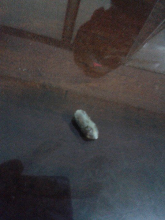
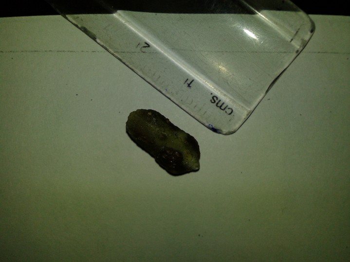
14mm Renal Caliculus removed nonsurgically at Dr SHAH’s Homoeopathy14mm Renal Caliculus removed nonsurgically at Dr SHAH’s Homoeopathy
For further reading and details on renal stones/urolithiasis/kidney stones click on the following link:
When solids and particulate metabolites or salts starts depositing at any level of urinary collecting system it forms renal stones or urolithiasis
Urolithiasis is more common in males than in females
Increased concentration in urine, of the constituents of stones is associated with stone formation.
1) Calcium Oxalate/Phosphate Stonescomprises 75% of every stones.
Its observed that almost 50 % of cases of Calcium Stones that have idiopathic hypercalciuria and doesn’t have hypercalcemia.
10 % of cases calcium stone cases have both hypercalcemia and hypercalciurea
5% have Enteric(4.5%) or Primary (0.5%) hyperoxaluria
20% have hyperuricosuria
15-20% have unknown metabolic anomaly
2) Struvite Stones (Magnesium, Ammonia, Calcium, Phosphate) usually due to renal infections comprises off 10-15% of all stones.
3) Uric Acid Stones comprises 6% of all stobe cases 50% of which are associated with hyperuricosuria and/or hyperuricemia and 50% are of idiopathic origin.
4) Cystine Stone comprises only of 1-2% of all the cases
5) Other and unknown types of stonescomprises of upto 10% of all the cases
An organic matrix of mucoprotien is present in all above all types of stones which acts as an additional binding matrix and it makes up almost 2.5 % of weight of all stones.
Renal caliculus, while selecting homoeopathic remedy for Kidney stones it is important to make note of all accompanying symptoms, like type of pain , character of urine, and if there are gravels in urine then its colour shape size should be noted, also other miasmatic and constitutional background should be evaluated for proper similimum remedy selection.
Risk factors
Increases Risk:
Dehydration, Low water intake, excess grapefruit or apple juice intake soft drinks containing phosphoric acid especially areated drinks.
Excess red meat consumption increases concerntration of certain sulfurous amino acids like cystiene and methionine which acidifies urine , decreases citrate excretion through urine and increases excretion of calcium and uric acid through urine thus increasing risk of renal stones.
Calcium suppliment tablets and VitaminD suppliment for long term may cause Calcium Stones
Studies show that low dietary calcium promotes calcium stones:- Unlike supplimental calcium , Natural dietary calcium actually protects you against renal stones. As it binds with ingested oxalate from food in gastro-intestinal tract to form calcium oxalate and thus reducing its absorption and inturn reducing oxalate concentration in urine. Oxalate is 15 times more potent in forming renal stones compared to increased levels of calcium in urine.
Sodium and fluoridated water increases urinary excretion of calcium thus increases risk of stone formation.
Low urinary excretion of citrate
Decreases Risk:
Unlike supplimental calcium , Natural dietary calcium actually protects you against renal stones. As it binds with ingested oxalate from food in gastro-intestinal tract to form calcium oxalate and thus reducing its absorption and inturn reducing oxalate concentration in urine. Oxalate is 15 times more potent in forming renal stones compared to increased levels of calcium in urine
Studies show that low dietary calcium promotes calcium stones.
Magnesium inhibits stone formation.
Patassium promotes urinary excretion of citrate and citrate inhibits crystallisation of calcium and thus reduces risk of urinary calcium stones
Commonly Used Homoeopathic Remedies For Renal Caliculus
Renal caliculus, while selecting homoeopathic remedy for Kidney stones it is important to make note of all accompanying symptoms, like type of pain , character of urine, and if there are gravels in urine then its colour shape size should be noted, also other miasmatic and constitutional background should be evaluated for proper similimum remedy selection.
Berberries Vulgaris sensation of retention of urine and urge to pass it again soon after passing, patient is thirstless, bright red mealy sediments, bubbling sore sensation in renal region, pain in urinary bladder region, pain radiating from lumbar region to pelvis and down to lower limbs while passing urine, burning in urethra even when patient is not passing urine, feequent micturation, well indicated in patients with renal caliculus with gouty diathesis and elevated uric acid levels and urate crystals in urine. Works well in patients having glomerulonephritis along with Renal Caliculus.
Hydrangea Arborescens white amorphous salt in urine giving urine a turbid colour, white amorphous salt in urine deposits in form of gravel. Sharp pain in loins especially in left side with unquenchable thirst for large quantity water, unlike berberries vulgaris which has thirstlessness. Well indicated in patients with enlarged prostate and also having Renal caliculus.
Cantheris In patients with bleeding due to renal stones, heamaturia and Nephritis, Intolerable constant urge to pass urine with paroxysmal acute cutting and burning from lumbar region to pelvis in front, urinary tract infection due to stone initially causing membranous scales giving appearance of bran in water when passed profuse urine and later on urine mixed with blood passes drop by drop jelly like shreddy urine , membranous scales giving appearance of bran in water, infection of urethra due to damage by renal stone causing irritation in urethra increasing sexual desire.
Sarsaparilla Scanty slimy flaky sandy and bloody urine, gravels in urine that passes in thin stream, this medicine should be thought of when sand-like gravels seen on daiper of children and they are cranky before and during passing urine, Kidney stones on right side.
Pareira Brava/ Condodendron Tormentosum great tenesmus while passing urine, constant urge to pass urine but has to strain much while passing has to bend down on his knees and press head on floor in front to pass urine causing pain to radiating till his thighs, Urine dribbles after micturating, pain in glans penis, renal stones in patient with gonorrhoeal urinary tract affections.
Borax Small red particles in urine and on daiper of child who cries while passing urine, hot smarting pain in urethra.
Solidago Redish brown thick sediment gravels with offensive albuminuria , pain in renal angle radiating forward and downwards towards abdomen and pelvis.
Silicea its a wonderful homoeopathy know for its ability to expel foreign particle from body, for which it is also termed as surgeons knife, Red or Yellow sedimentation prostorrhoea due to tenesmus in patients with retention of urine due to renal caliculus
Lycopodium Heavy Red Sand Particle in urine, pain in right hypochondrium and right lumbar region , renal stones in old men with slow feeble urine with stangury, tenesmus and retention of urine.
Calcarea Carbonica Calcium oxalate crystals in urine, sour fetid smelling urine with dark brown colour and white sediments and haematuria.
For further reading and details on renal stones/urolithiasis/kidney stones click on the following link:
Vitamin B 12 also called Cyanocobalamine, has the largest molecule compared to all other vitamins.
BIOCHEMISTRY
Vitamin B12 is plays a major role to synthesis
1) Methylmalonyl CoA to Succinyl CoA
2) 5-Methyltetrahydrofolate and Homocysteine to Tetrahydrofolate and Methionine
VITAMIN B12 IS NECESSARY FOR
Myelinogenesis , its co-substrate in various cellular reacations involved in methylation synthesis of nucliec acid and neurotransmitters, active metabolites of vitamin B12 are required for methylation of homocystine which is required in formation of methionine, which is required for metabolism of many monoamine neurotransmitters, thus it plays a mojor role in proper nervous system formation and its functioning.
DNA synthesis thus helps in proper maturation of neucleus and cells.
Metabolism of amino acid and fatty acids in cells.
DEFICIENCY OF VITAMIN B12
Its deficiency can cause severe damage to nervous system
As it is essential for each and every cells, its long deficiency may cause damage to organs and may result other metabolic disorders.
General symptoms it present on slightest decrease below its normal range are.
General Weakness
Dyspnoea on Slightest Exertion
Fatigue
Lethargy
Pain in Limbs
Depression
Somnolence
Insomnia
Weak memory
Mania
Depression
Heachces
As it is essential for each and every cells, its long deficiency may cause damage to organs and may result other metabolic disorders
ESTIMATED DAILY REQUIREMENT OF VITAMIN B12
Infants upto age of 12 months its from 0.4μg/day to 0.5μg/day
Children upto 13yrs of age is 0.9μg/day to 1.8μg/day
Person above 14yrs of age is 2.0μg/day
Pregnant Women is 2.6μg/day
Lactating Women is 2.8μg/day
Infants upto age of 12 months its from 0.4μg/day to 0.6μg/day
DIETARY SOURCES OF VITAMIN B12
Meat and Fish are rich source
Except for Arachea and Bacteria no other In cellular life forms has genes to express protiens that can produce or synthesis vitamin B12 De Novo.
Vit B12 is absorbed in small intestines and there are bacteria which produce Vit B12 in colon so , even though its produced by microbial flora of intestine of humans it is difficult to absorb.
Ruminating vegetarian animals like cow buffalo etc they can consume their gut Vit B12 produced by gut bacteria when they bring back up the food to chew and deglute again, also they pick it up when they graze grass along with the soil at roots of grass they get ample of bacteria which provide them with Vit B12.
There are very few vegeterian dietary sources of Vit B12 and also they are very low in Vit B12 quantity,
So vegetarian are more prone to Vit B12 deficiency,
Major reason for deficiency is insufficient dietary intake of Vit B12.
In many cases malabsorption and lack of factors required for binding Vit B12 causes its deficiency,
Infants feeding on mother with low vitamin B12 may develop deficiency.
In vegan population incidence of vitamin B12 deficiency is estamited to be upto 80%
SOURCES OF VITAMIN B12 FOR VEGETARIANS
So for vegetarian it is recomended to consume commercially available fortified food with Vitamin B12
Fortified oats
Dairy products like
Milk
Paneer
Curds
Nutritional Yeast
Probiotics
Cheese
Fortified cereals
Eggs
Its is strongly recomended for vegetarians that if they have developed deficiency then they should not depend only on dietary sources but also start Vitamin B12 suppliments either oral or injectable whatever recomended for each individual case.
Homoeopathic medicines helps regularise weight of a person as per constitution and requirements of an individual.
For optimal realisation of Homoeopathic medinical effects the medicines needs to be selected as per the constitution with special emphasis on physiological imbalances and any other underlying pathological factors needs to be ruled out.
If not selected in accordance with the homoeopathic principles, the medicine might not yield desired result.
Similarly if selected in accordance with Homoeopathic principles it will not only help in weight gain but also resolve any other underlying abnormalities found in proper physiological functioning of body. As the medicines work via improving constitution of a person.
Below are simplified guidelines to select few of the generally used common medicines which are easy to select that most of the instances are hard to fail and can help gain weight in individuals with below normal weight.
Abrotanum especially suited to person with Protein Energy Malnutrition, in ascending type of Marasmus, pot belly with lean thin arms and limbs, in person who eats well but still loses flesh Kwashiorker Protein Energy Malnutrition PEM , in person with Gouty Diathesis who lose muscles, in person who lose weight due to chronic Diarrhoea dysentery, linteric stool with visible undigested food particles in stools.
Alfa Alfa general homoeopathic tonic to improve digestion promote protien absorption and metabolism.
Ashwagandha / Withania promotes testosterone secretion thus promoting protien metabolism improving muscle mass and reducing fats
Natrum Mur weak lean malnourushed emaciated person with unhealthy skin and earthy complexion, irritability and depressive temperament causing weight loss.
Tuberculinum weightloss due to past or present tuberculosis
Phosphorus lean thin pale person with history of tuberculosis and sensitive to cold.
Arsenicum Album typically suited in patients with Obsessive Compulsive Disorders, bulemia nervosa, person stops eating due to fear fright and anxiety of death, disease or obesity.
Chelidonium Majus weight loss due to sluggish liver function
Lycopodium low body weight or weightloss due to liver disease or sluggish liver function.
Pulsatilla in women with depressing emotion weeping desposition pale fair complexion with tendency towards hyperacidity and tendency to catch cold easily.