
Anaemia can be defined as decreased haemoglobin counts or reduced red blood cell counts or reduced oxygen carrying capacity of blood, due to “loss of” or “abnormality of” red blood cells or haemoglobin.
Normal Heamoglobin Counts
- 6 months to 5 years of age > 11g/dl
- 5 years to 12 years of age > 11.5g/dl
- 12years to 16 years of age > 12g/dl
- Adult Females (non-pregnant) > 12g/dl
- Adult Females (pregnant) > 11gm/dl
- Adult Males > 13g/dl
CAUSES OF ANAEMIA
- Blood losss
- Excessive Red Blood Cell destruction
- Heamoglobinopathies
- Hypovitaminosis B12
- Hypoferremia
- Anaemia of Chronic diseases
- Autoimmune haemolytic anaemia
- Inflamatory bowel diseases
- Hypervolemia or water retention due to sodium or other salts.
- Genetic hereditary conditions like Thalasemia
- Certain cancers
- Kidney diseases
- Reduced erythropoetin production
- Excessive RBC destruction
- Impaired RBC production
- Certain infections like malaria which causes RBC destruction.
- Certain drugs which causes RBC destruction eg. Quinine causes chinchonism.
- Bone Marrow lesions and pathologies
- Etc.
CLASSIFICATION OF ANAEMIA
There are many types of anaemias. It can be broadly classified into 7 categories depending upon their causes
Anaemia due to
- Blood Loss
- Hemolysis
- Impaired or abnormal Erythropoesis
- Hypervolemia
- Chronic Diseases
- Nutritional deficiency
Based on RBC morphology it can be classified into 3 groups
- Microcytic
- Macrocytic
- Normocytic
FEW COMMON and RARE TYPES OF ANAEMIA
- Iron Deficiency Anaemia
- Aplastic Anaemia
- Megaloblastic Anaemia
- Pernicious Anaemia
- Sideroblastic Anaemia
- Autoimmune Hemolytic Anaemia
- Myelodysplastic Syndrome
- Thalasemia
- Fanconi Anaemia
- Congenital Dyserythropoetic Anaemia
- Daimond-Blacfan Anaemia
- Myelopthisis
- Anaemia of Prematurity
- Erythroblastopenia or Pure Red Cell Aplasia
- Hereditary Spherocytosis
- Hereditary Elliptocytosis
SYMPTOMS
- Weakness
- Lethargy
- In children it affects growth in general
- Somnolence, Drowziness in day time
- Disturbed sleep at night
- Pallor, general pale appearance of skin, mucous membranes and eyes.
- Dyspnoea on Exertion.
- Reduced Immunity, tendency to catch infections and slow recovery and healing.
- Bodyaches
- Cyanosis in severe cases
- Palpitations
- Tachycardia
- Low blood pressure
- Chest pain
- Depression
- Craving for indigestible things , PICA
- Cold clammy extremities
- Oedematous swelling of extremities, dependent oedema
- Angina or cardiac failure in severe cases
- Will impact general growth and repair of all the vital organs and tissue of the body.
HOMEOPATHIC MEDICINES FOR ANAEMIA
Depending upon the cause of anaemia and general constitution of the patient, one of the following medicines may be called for duty by a homeopathic physician.
- Ferrum Metallicum
- Ferrum Phosphoricum
- Cinchonna Officinalis
- Natrum Muriatic um
- Arsenicum Album
- Abrotanum
- Hamamelis Verginiana
- Pulsatilla Nigricans
- Janosia Ashoka
- Crotalus Horridus
- Lachesis
- Acidum Phosphoricum
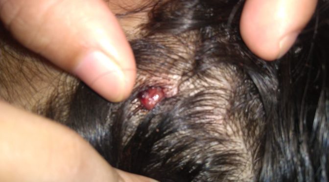

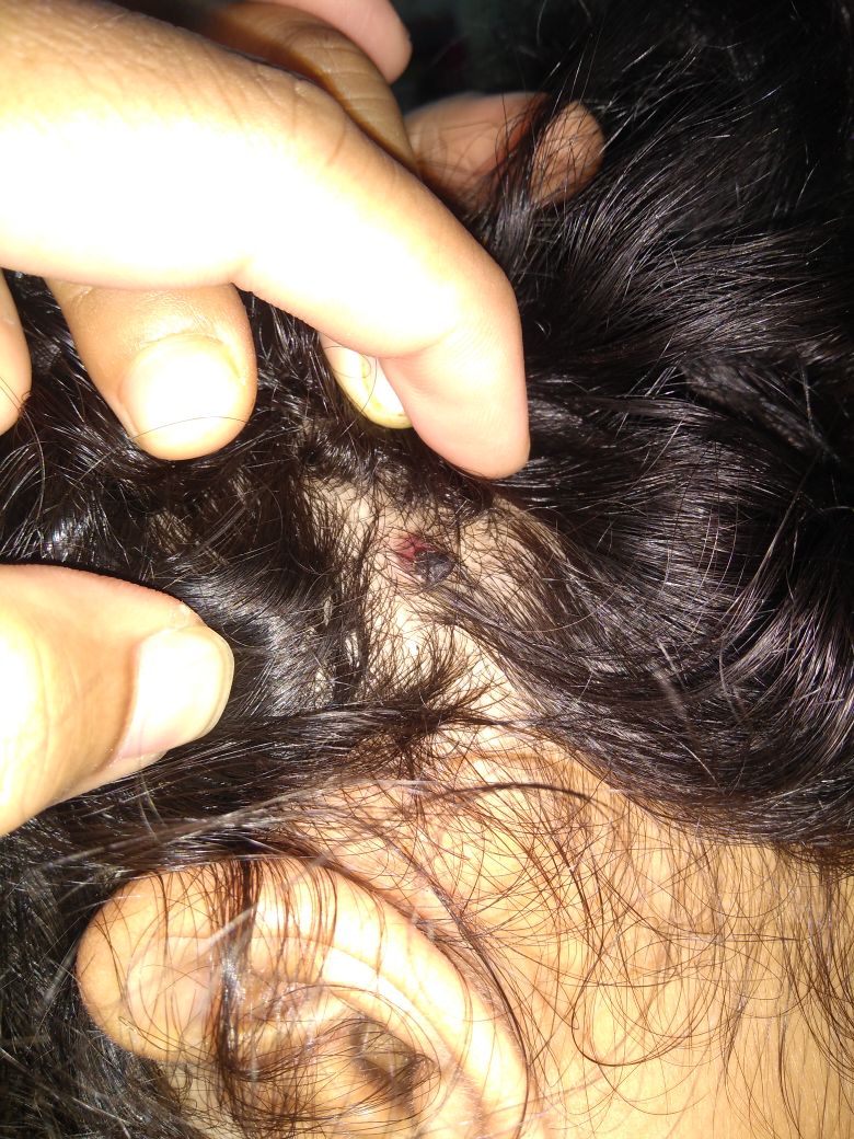


 BEFORE TREATMENT
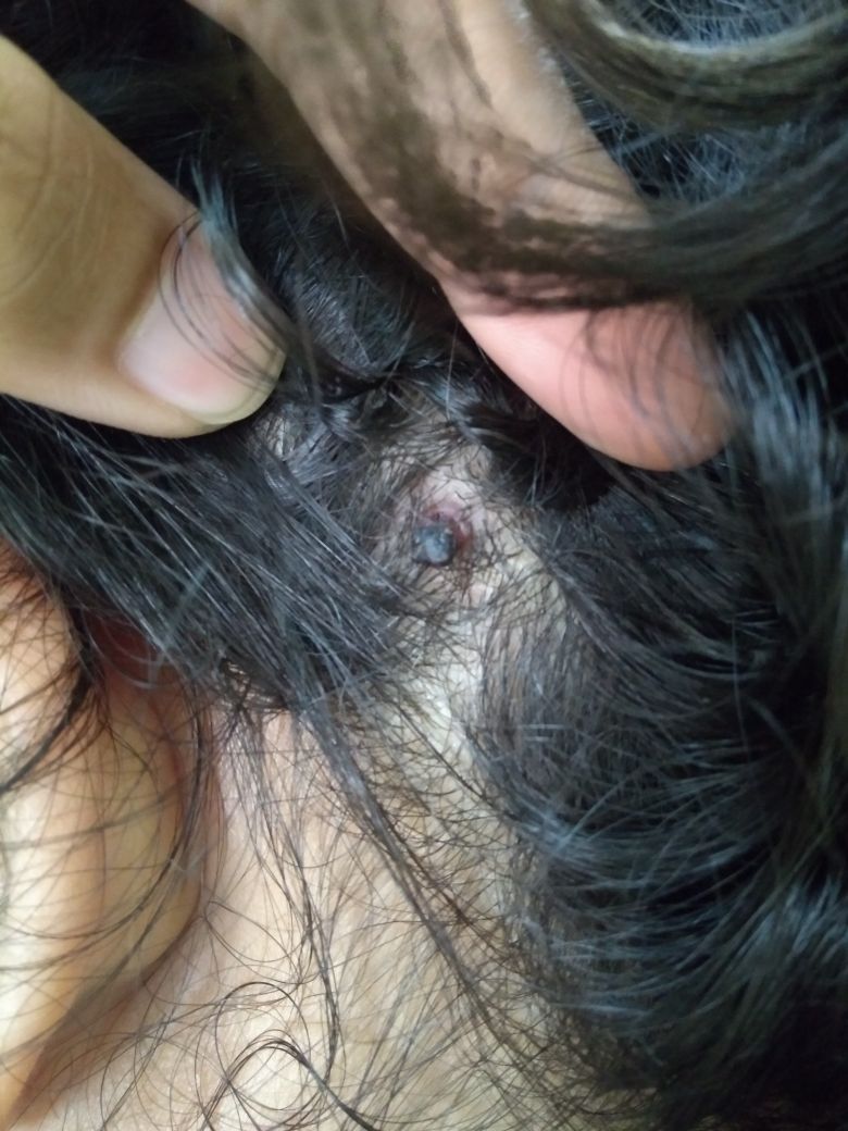
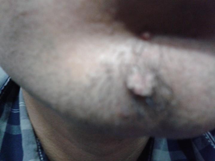
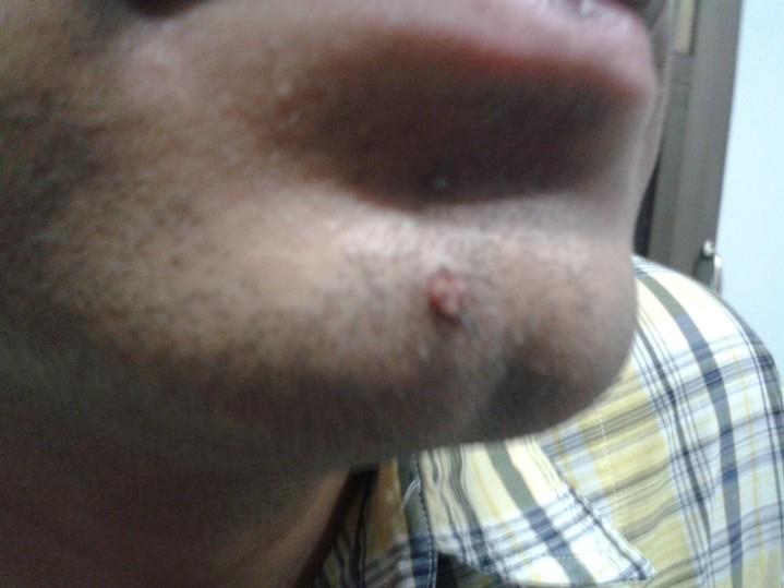 DURING TREATMENT
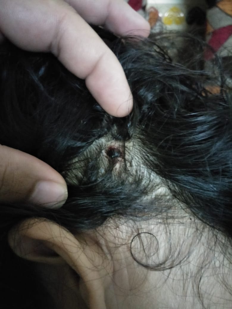
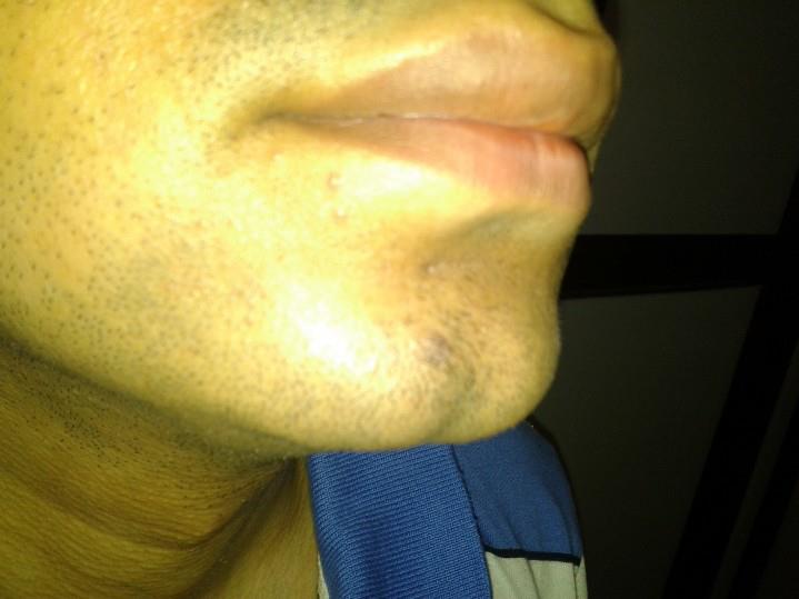 35 DAYS AFTER TREATMENT
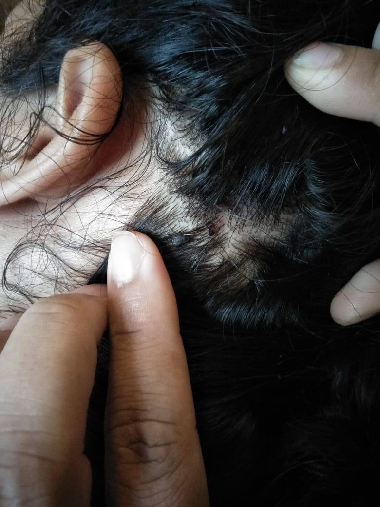