PYOGENIC GRANULOMA also called GRANULOMA TELANGIECTATICUM or LOBULAR CAPILLARY HEAMANGIOMA is relatively a common benign vascular lesion of skin and mucosa and appears as an overgrowth of tissue.
It is usually a small, round, bloody red in color.
They are also known as Lobular capillary haemangioma or granuloma telangiectaticum.
They grow rapidly at first and then remain a constant size, they rarely exceed 1cm in diameter.
What causes Pyogenic Granuloma?
The following factors have a possible role in their development
Some cases develop at the site of a recent minor injury, such as a pin prick.
Staphylococcus aureus is frequently present in the lesion.
Hormonal changes in the body during pregnancy can also be the cause of Pyogenic Granuloma.
Certain drugs and medications are known to cause pyogenic granuloma
SIGNS AND SYMPTOMS
The appearance of Pyogenic Granuloma is usually a small red, round, oozing and bleeding bump.
Younger lesions bleed easily because they are rich in blood vessels. These lesions can be painful.
The lesions arise on various regions of the body such as eyelids, face, tongue, lips, arms, hands and feet.
EYELID: The condition gives rise to reddish pink vascular bumps over the eyelid and is said to be caused due to a rich blood supply from conjunctiva.
TONGUE: These growths arise on lateral side of the tongue, it may arise due to trauma from dentures.
LIP: Arises as a solitary growth on the lip.Growth generally appears on the lower lip.
FINGERS:The growths are common on hands and fingers.
FACE: Growths arising on face are generally multiple in number and looks like small red or reddish purple lumps
FEET: These bumps arise in the nail bed of a toe with an accompanying ingrown toe nail.
DIAGNOSIS
Pyogenic Granuloma can be diagnosed based on its appearance.
HOMEOPATHIC MANAGEMENT
Commonly Used Homeopathic Medicines for Pyogenic Granuloma
SYPHILLINUM
Syphillinum is mostly indicated in case of pyogenic granuloma, where the patient presents with abnormal growths with pain. Indicated in case of chronic erruptions. Suited to person with pale and fine textured skin, who are slender. Presents with marked prostration and debility.
HEPAR SULPH
Hepar sulph is indicated in pyogenic granuloma. Specially suited to lymphatic and phlegmatic individuals. Excessive sensitiveness of parts is a leading indication. Chilly sensation. Indicated in csae of vascular lesions of the skin, they are bright red in colour which tend to bleed.
SILICEA
Silicea is mainly indicated in cases like pyogenic granulomas. Patient presents with vascular lesions of the skin where the suppurations continue and wound refuse to heal. Discharge is thin bloody. The lesion are benign and granulomatous.
THUJA OCCIDENTALIS
Thuja is indicated in case pyogenic granuloma as it is used in bleeding growths, spongy tumours, warts. Thuja has main action on the skin. The lesions have tendency to bleed easily and presents with pain. Complaints worse at night.very sensitive to touch.
RUTA GRAVEOLENS
Ruta is indicated in case of pyogenic granuloma. The patient presents with small rounded vascular lesions which tend to bleed easily. Very anxious and low spirited with mental dejection. Presents with neuralgia, burning stinging pain.
CALCAREA FLOURICA
Indicated in case of pyogenic granuloma. Vascular lesions which tend to bleed and are painful. Indicated in abnormal grwoths. There is great depressio . Sensitiveness to cold. Mostly indicated in blood tumors.
Homeopathic Approach in case of Pyogenic Granuloma
In Homeopthy we not only consider the signs and symptoms of the patient but treat the patient holistically, based on individualisation.
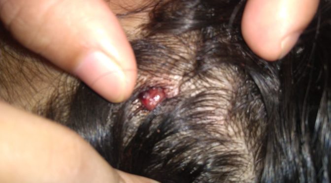
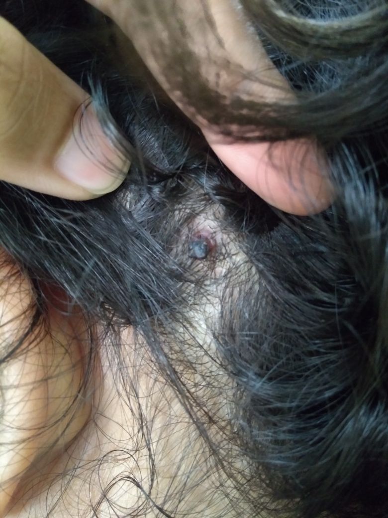
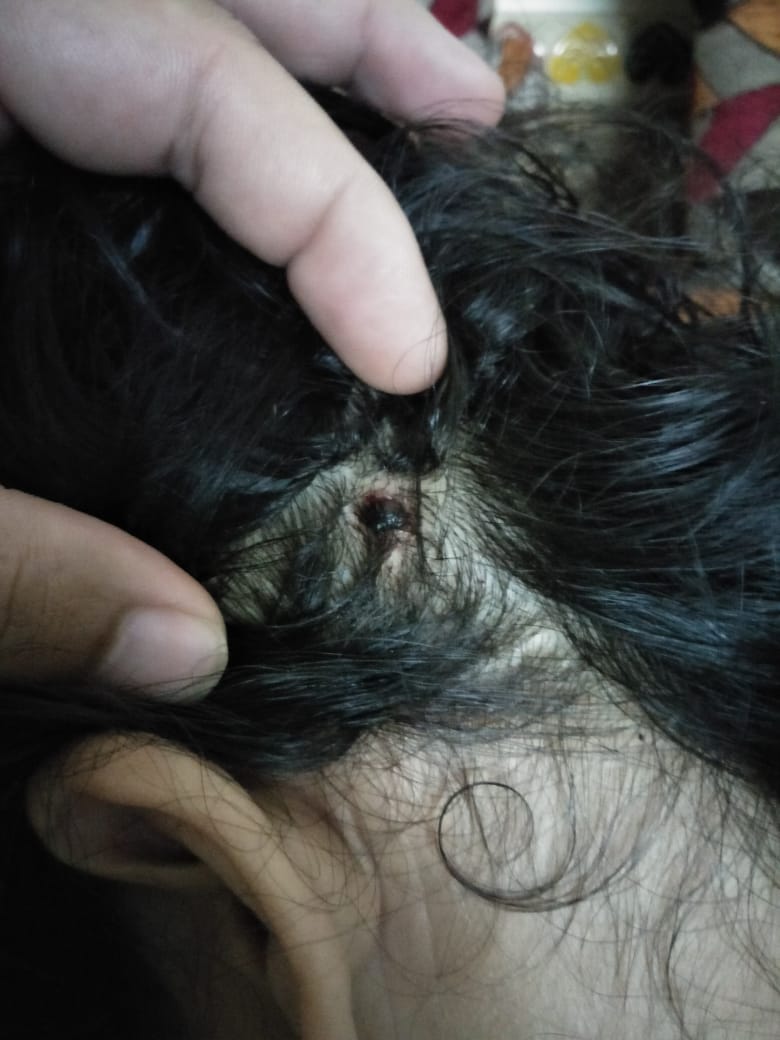
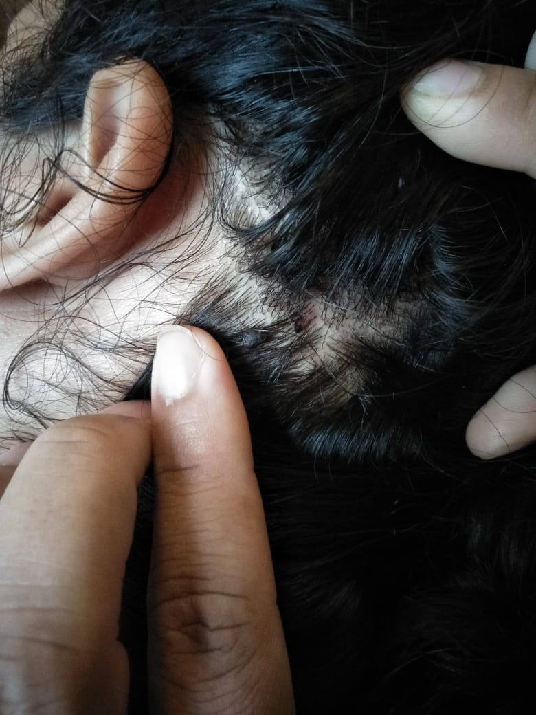
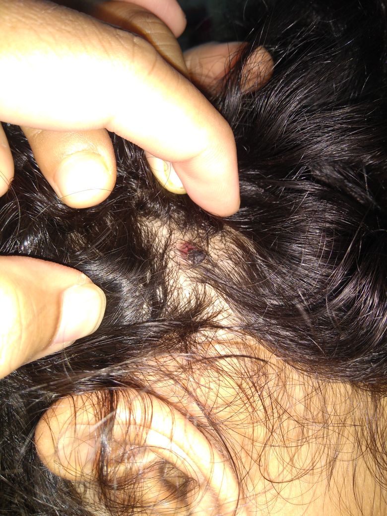
Similarly considering the symptoms-similarity, individuality and total of the patient, below is an example of one of our cases of pyogenic granuloma at Dr SHAH’s HOMOEOPATHY this case responded well to homeopathic medicine Syphillinum and got completely resolved with selected potency of 200C of the said remedy.
Pyogenic Granuloma Before treatmentDuring Homeopathic treatment Pyogenic Granuloma becoming dark and dryDURING HOMEOPATHIC TREATMENT PYOGENIC GRANULOMA BECOMING DARKER DRIER AND SHRINKING IN SIZEDURING TREATMENT – SEE THE LARGE FRAGMENT OF PYOGENIC GRANULOMA HAS GOT DETACHED AND HAS INTERMINGLED IN HAIR NEAR INDEX FINGERDuring Homeopathic Treatment at Dr Shah’s HomeopathyPyogenic Granuloma resolved after detaching and shrinking completely now only a small dark spot can be seen at the site.Pyogenic Granuloma- After Treatment – Cured completely after Homeopathic Treatment By Dr DEEPAN P SHAH at Dr Shah’s Homoeopathy.
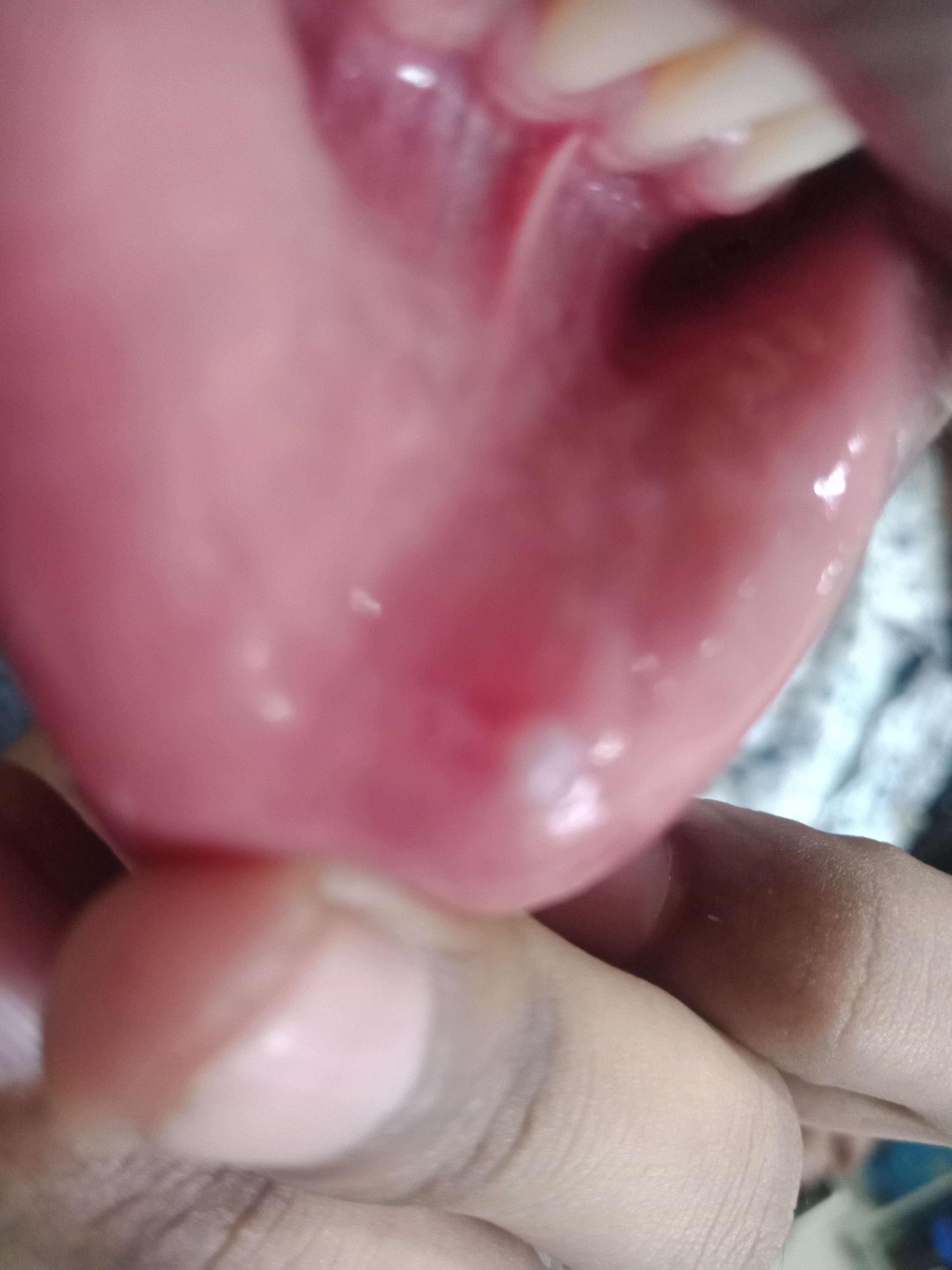
Oral Mucocele is a cyst formed due to collection of salivary gland secretion.
It is pearly translucent occasionally with bluish discoloration usually measuring from 1-8mm in diameter. The deeper cysts may appear as a nodule and may not have translucent pearly appearance as in superficial cyst.
It appears on lower lip on sides.
When it appears on floor of mouth below tongue its termed as ranulla.
They are painless most of the time.
It usually lasts for few days to years.
It may frequently rupture and reappear again at same site.
Ruptured Salivary Gland Duct this results into mucous extravasation phenomenon.
Obstructed Salivary Gland Duct this results into mucous retention cyst.
Rupture or Obstruction can be due to
Infection that can potentially obstruct or damage the salivary gland and its duct. which can be due to many factors most common being poor oral and dental hygiene.
Injury on lower lip which can damage salivary gland duct may result innto oral mucocele tje most common form of injury is due to bitting lips by one own teeth.
Tumours that may obliterate obstruct or rupture or damage salivary glands and its ducts in any form may result in to mucocele in oral mucosa
HOMEOPATHIC TREATMENT FOR ORAL MUCOCELE
The underlying cause needs to be evaluated, exclude the probability of infection, malignancy. Patient needs to be guided for proper oral hyegiene, any habbit like bitting teeth should be avoided.
Following are the common Homeopathic medicines used in treatment of Oral Mucocele
Silicea
Thuja
Variolinum
Calcarea flourica
Mercurious Solubilis
Syphyllinum
Antimonium Tartaricum
Antimonium Crudum
Apis Mellifica
Ranunculus bulbosus
Selection from the above medicines should be strictly done as per symptom similarity basis under consultation of homeopathic practitioner.
Nipah Virus Homeopathy Controversy – Does Homeopathy Really Have Prevention or Cure For Nipah Virus Infection?
Nipah Virus Homeopathy controversy has been gaining momentum since few days with many claiming that the claims of homeopathy are false, so whats the truth is lets have the deeper understanding about it.
Nipah Virus outbreak in state of Kerala first noted on 19th of May 2018 and since then the government authorities are on alert and to contain the infection and avert the potential epidemic about 230 doubted cases have being tested of which total 18 have turned out positive of which 16 have already lost their life. which shows that mortality rate of current outbreak is very high compared to average of all past outbreaks one can say almost 100% fatality rate of the current outbreak
THE NIPAH VIRUS HOMEOPATHY CONTROVERSY
Homoeopaths have come out with the medicines that can be indicated or applied in subjects of Nipah Virus Infection after proper homeopathic evaluation. To which believers of orthodox wide spread conventional mode of treatment, the allopathy, have come up with a very rebellious mood as usual claiming that homoeopathy cant work in Nipah Virus Infection and rebuting our claims even without giving a thought find out its practicality.
Homeopathy has since long came in for the disposal of mankind whenever there were epidemics and outbreaks of disease and allopathy had failed.
EXPLANATION – WHETHER HOMEOPATHY HAS MEDICINE FOR NIPAH VIRUS INFECTION OR NOT
Now the question arises right now is of the current outbreak of Nipah Virus Infection and its homeopathic treatment whether homeopathy has medicines for prevention and/or cure of Nipah Virus Infection or not.
Homoeopathy is based on principle of “likes cure likes” its called the principle of similars ( just like vaccines ) ; where as the Allopathy is based on dissimilar sufferings and so aptly the name allopathy to this system was given non other than by the Founder of Homeopathy Dr Samuel Christian Fredrich Hahnemann and since that time Allopath’s opposition to this system Homeopathy is continuing.
In homeopathy we do not treat “the disease” but we treat “the person in disease” – Various Disease causing agents have different type of virulence, each which very specific set of characters, now our immune system responds in various ways to various antigens and also its very much subjective in various individuals. So in homeopathy we first take into consideration the type of response of immune system and what it has to express and how its responding to this antigenic stimulus as it is the one which is going to fight out the disease.
So in homeopathy we do not have name of the disease and its medicine but we have the set of symptoms that our immune system is expressing and its matching remedies. So its based completely on symptom similarity principle with which homeopathy since ages have cured many so-called incurable conditions because we believe in treating the Person in Disease by understanding what its immune system is expressing and trusting and strengthening it so that it can drive out the antigen with its dynamic, intelligent, automatic, autocratic, powerful nature of force which has helped life forms to evolve from unicellular to various life forms so different from each other and with different abilities.
In Homeopathy rather than hitting the virus or the antigen directly with noxious agents and damaging the system even further we simply trust the unquestionable power and ability of our immune system and try to strengthen it by stimulating it so that it can drive the virus out so homeopathy shows better success rate in treating viral conditions compared to allopathic mode of treatment.
Now in Nipah Virus Infection in past outbreaks we have seen that there are IgG and IgM antibodies against Nipah Virus have been formed in individuals that have survived the infection. so the immune sytem of many is strong enough to fight out the virus and survive.
CONCLUSION TO THE CONTROVERSY OF NIPAH VIRUS HOMEOPATHY
So homeopathy has a role in the Nipah Virus Infection disease and homoeopathic medicines if selected as per symptoms of patients affected by the disease and principle of organon of medicines should help the Nipah Virus Infected subjects to recover from the infection completely and also prevent the condition as mention in principle of Genus Epidemicus.
Journalists and critics should responsibly first understand homeopathy and its principles which are quite different than allopathy before jumping up on conclusion that homeopathy has no solution, similarly homeopathic doctor or association should be very careful mentioning indicated medicines and should not make any unproven claims of sure cure or sure prevention and should be very clear that the medicines are only indicated as per homeopathic principles.
So health authorities with unprejudiced and unpolitically motivated mind should come forward and consider the goods of the homeopathic system of medicines which the homeopathic system has delivered since ages and should apply in the preventive medicine for nipah virus and treatment of Nipah Virus Infection to confirm and perfect the homeopathic community claims, Especially when where allopathy is absolutely failing!
Neurofibromatosis is syndrome of Phakomatosis or Neuro-Occulo-Cutaneous type of Autosomal Dominant Genetic Anomaly. It can be hereditary germline change or acquired during early developmental stage, affecting structures of embryonic ectoderm, where in, there is mutation or rarely somatic mosaicsism(RASopathy) of NF1 gene or its alleles, present on Chromosome 17 and is responsible for synthesis of protein neurofibromin which regulates cell proloferation and differentiation which hampers tumour suppressor activity giving rise to tumours of nervous system.
TYPES OF NEUROFIBROMATOSIS
NEUROFIBROMATOSIS TYPE 1.
It is considered to be syndrome falling under category RASopathies. It affects supporting structures of Peripheral Nervous System. This type rarely show any severe complication other than cosmetic and compression complaints.
NEUROFIBROMATOSIS TYPE 2.
Involves Schwaan cell of Cranial nerves especially of Vestibulocochlear nerve that is 8th cranial nerve or CN VIII causing bilateral acoustic Schwannoma causing hearing loss. It shows many complications of cranial nerves and brain stem. It can be disabling and life threatening. Severeity varies in different individuals depending on the way in which the disease assumes its course in different individual.
SCHWANNOMATOSIS.
In this type Schwann cell tumours are found in Peripheral and Spinal nerves. It can be very painful and disabling.
SYMPTOMS OF NEUROFIBROMATOSIS
Multiple upto six or more “cafe au lait” spots more than 5mm in diameter is one of the diagnostic sign which are also present in Mc Cune Albright Syndrome.
Freckles generalised dispersed throughout body increasing simultaneously with neurofibromas.
Excessive folds of skin especially in underarm region with freckles.
More than one overgrowths in iris of eye.
More than one Neurofibroma.
Spinal growth and curvature anomalies.
During early stages of development in children it may show Learning and Behavioral problems
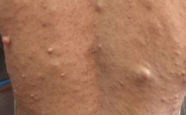
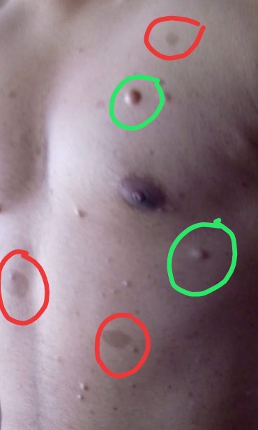
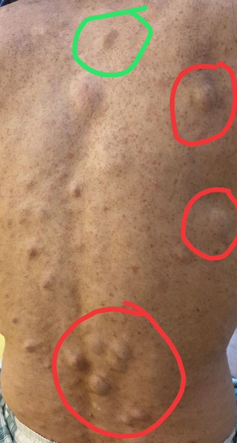
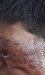
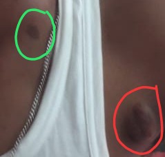
Cafe au Lait spots and neurofibroma in patient with NF 1)Neurofibroma marked in Green Circles 2)Cafe au Lait spots marked in Red CirclesNeurofibromatosis patient with classical presentation of Cafe au Lait Spot, Neurofibromas and extensive freckles on back. 1)Café au Lait spot – dark brown spot marked in green circle 2) Neurofibroma – nodules marked in red circles 3)Freckles – Extensively scattered, small dark dots like pigmentation throughout the back Above three are common findings in patients with Neurofibromatosis.Plexiform Neurofibroma of eyelidsCafe au Lait spots and neurofibroma in case of NF. 1)Neurofibroma in Red Circle 2)Cafe au Lait Spot in Green circle
DIAGNOSIS OF NEUROFIBROMATOSIS
HISTOLOGY helps to confirm neurofibroma
SLIT-LAMP TEST
GENETIC TESTING helps confirm the diagnosis.
EEG, MRI, CT SCAN AND MRI helps us to assess tumour and its influence on surrounding tissue
Sphenoidal dysplasia on imaging, is one of the major finding in diagnosis of NS1
Sun Stroke or Heat Stroke is a fatal condition caused due to exposure to excessive heat of Sun
Its also called Non Exertional Heat Stroke (NEHS).
And Exertional Heat Stroke (EHS) is the condition where it is caused due to excessive physical exercise.
As the summer approaches, so does the increase in number of sun stroke or heat stroke cases. In summer of 2015 more than 2500 people died in India. Many of them were from metro cities having best medical facilities. Many more world wide are losing their life to this fatal avoidable medical emergency.
RISK FACTORS of SUN STROKE or HEAT STROKE
Summer season
Heat waves
Humid Climate
Eecessive physical exertion
Pre-existing acute infection in body
Heart diseases
Skin ailments
Allergies
Autoimmune conditions
Beta-blockers
Diuretics
Alcohol
Drug Abuse
Smoking
Diabetes
Hypothyroidism
Other Chronic Diseases
Children and Elderly subjects
It is a medical emergency and requires immediate medical attention, the sooner its detected the better the prognosis and lesser the long term consequenses, yes heat stroke does lead to consequences even after patient recovers from convalscence which was best understood after 1995 Chicago heat wave, when 50% died within a year, and many has disabilities and impairment making them disabled and dependent.
SIGNS and SYMPTOMS of SUN STROKE or HEAT STROKE
Hyperthermia, Increased body temperature usually above 105°F or 40.6°C
No Perspiration/ but in Exertional Heat Stroke patient has much perspiration.
Dry red skin / damp skin if not exposed to heat of sun for prolonged hours and or was in humid climate.
Symptoms might not be much evident if patient is in sleep.
The disease soon progress to show severe sypmtoms like unconsciousness, seizures(usually in children).
Rhabadomylosis
Organ failure, kidney and heart in particular
And death ensues.
It is important to judge the condition earliest but its also difficult as initially patient doesnt realise that its a sun stroke and carries on with the mild discomfort he is experiencing and soon symptoms starts progressing.
So its necessary to educate people about its causes, how to prevent it, occurence of symptoms and what to do at first if doubted.
PREVENTION of SUN STROKE or HEAT STROKE
Dont’s to prevent Sun stroke or heat stroke avoid following things
Exposure to heat for long
Excessive exercise or exertion especially in hot climate especially military personals, athletes, fire fighters and other straineous activity workers in hot climate needs to take extra precautions.
Sitting in car without aircondition it frequently happens when car is parked. Even when out side temperature is around 20-21°C the temperature inside car can reach upto 49°C , without the aircondition and when parked even with slight window open.
Leaving behind children and elderly in parked car.
If the climate is too hot and humid and you are with children dont neglect or forget the fact that they are more prone to the condition comparatively, and u may subject them to it and this happens too frequently which is why its now commonly termed by laymen as “Neglected Baby Syndrome” a non medical term famous tailored by laymen which discribes the situation and not the disease.
Avoid stimulants, smoking, alcohol and drugs in hot climates.
In acute infection or other acute illness avoid being in hot humid climate for too long.
In chronic ailments especially metabolic disorders or allergies or autoimmune conditions avoid prolonged exposure to heat or exertion.
If you are on beta-blockers or other diuretics, then avoid exertion or heat exposure.
Avoid heat exposure or exertion in heart or kidney disease.
Do’s to prevent Sun Stroke or Heat Stroke
Drink ample of water in short intervals of time even if u are not feeling thirsty, in hot climate, also note excess of water in electrolyte imbalanve may be counterproductive and dangerous.
Keep sipping commercially available isotonic sports drink.
Maintain you urine colour light, dark urine colour shows signs of lack of water, urine colour is better indicator than the feeling of thirst.
Eat lots of salads and be more on liquids if you in hot climate.
Wear loose, white, thin, clothing a cap or hat with ample ventilating holes and large brim helps protect from heat.
Keep searching shades and cool places and halt for a while if u are walking in hot summer sun.
Keep body dry keep wiping body sweat.
Block direct heat of Sun.
Know the causes, prevention, signs and symptoms and first aid of Sun Stroke or Heat Stroke
TREATMENT of SUN STROKE or HEAT STROKE
Patient should be first taken under proper medical observation as soon as possible when ever available.
First and foremost aim is to bring down body temperature and to hydrate the patient.
Mechanical means to cool the patient as soon as possible can be applied.
Remove all clothes of patient and wipe off all sweat and make his body dry.
Cool and dry climate should be provided airconditioned room preferably or highly ventilated cool dry climate.
Avoid cold wet cloth wraps generaly on whole body. It might act as insulation and prevent heat to escape out from the body. Instead use cold packs only on head, neck, torso and groins, let rest of the body be open.
Bathing the patient in cold water is a good idea. Immertion in too cold water water is also a good idea. Both of these were previously opposed, due to the doubts of vasoconstriction of superficial blood vessels, preventing heat to escaping out of body. However more practical evidence and study have proved it to be useful.
Hydration of patient should be done simultaneous. Excess of water in presence of electrolyte imbalance can make case worse, so isotonic sports drinks comercially available are good option.
Intravenous fluid infussion whenever required, keeping in chk the electrolyte imbalance and accordingly infusing the indicated IV Fluid.
CRP and other measures in complicated cases in hospital setup.
HOMOEOPATHIC MEDICINES for SUN STROKE or HEAT STROKE
General guidelines for Homoeopathic medicine section for sun stroke or heat stroke
While Selecting Homeopathic medicines for Sunstroke or Heat Stroke one needs to first understand that Heat Stroke is a fast progressive acute condition.It is caused due to excessive heat causing disturbed thermo-regulation of the body, with loss of water from body altering electrolytes and enzymatic processes of cellular respiration.
So one need to be very careful about related aspects that can not be neglected which are as follows:
Medicines needs to be fast and deep acting with more affinity towards CNS Heart and circulatory system along with urinary system.
Such cases demands frequent repetition of doses.
Type of thirst pattern of medicine and that in patient with sunstroke are of utmost importnce.
Skin symptoms with characteristic appearance and texture cant be neglected.
Also make note of the characteristics of patient’s tongue appearance and try to match with remedy
Characteristics of Urine and Urinary system needs to be evaluated with much attention.
Fever and headache pattern and other concomittants during fever like general condition, delirium level of consciousness etc are important.
Pulse pattern needs to be observed.
Neupsychiatric condition evaluation is must.
Acessesment of all other organs especially heart and kidney is necessary to rule out complications.
Such cases needs to be managed under highly trained medical staff.
Homoeopathic medicines can be administered along with regular medical supportive management.
Following are some Homoeopathic Medicines for Sun Stroke along with their indications.
Strictly to be taken under guidance and observation of qualified homoeopathic doctor and medical staff.
ACONITUM NAPELLUS –
Fever and Headache Acute violent sudden onset of acute heavy pulsating headache with sensation as if head floating in boiling water. Acute onset of violent fever with fear, fright and anxiety especially fear of death with mental restlessness. Complaints caused due to dry climate and extremes of heat or cold. Much indicated in very first stage of sunstroke, the sooner you give better the results. This remedy is indicated only in functional disturbances there is no evidence of aconite being useful where there are gross pathological changes also its effect is very short without any periodicity.
Thirst – intense thirst, drinks and vomits and declares himself that he will die.
Tongue -Dry red constricted tongue with white coating and tingling on the tip of tongue.
Urine – scanty, red, hot, painful.
GLONOINUM
A theraputically specific remedy for sunstroke in homoeopathic materia medica, which can be applied in general with much benefits in most of the cases of sun stroke.
Headache and Fever –Surge of blood to the head and heart. Aggravation due to exposure to heat of fire or heat of sun. Sensation of pulsation throughout the body. Throbbing headache with diziness and confusion, cannot recognise localities. Head heavy but cannot lay it on pillow as he cant bear any heat around head and also it aggravates throbbing rush of blood to head, visual disturbances during headache sees everything half light half dark with visions of sparkling light.
NATRUM MURIATICUM –
A known remedy for sun stroke and complaints due to heat of sun better given in biochemic form especial in children and female of pre-pubertal and early pubertal age affected with sun stroke effects in early stages. Throbbing blinding headache with sensation as if thousands of little hammers were knocking.
Thirst -unquinchable thirst for cold drinks, desires salt.
Tongue -Mapped tongue frothy coated tongue with cracked lips with numb sensation.
Skin – Greasy and Earthy texture , Blistering redness with much burning due to sun burns with dry sensation.
Urine – pain in urethra after passing urine, involuntary urination.
BELLADONNA–
Headache and Fever-Very well indicated when there is severe headache with throbbing heat especially in forehead and temples with vertigo and high grade fever and difficukty in breathing
Tongue – Dry, Swollen glazzy and Painful with red edges, Strawberry like appearance of tongue due to elevated buds and papillae of tongue,dry throat.
Thirst –Great thirst for cold water with nausea and vomitting and spasm of stomach, but thirstless during fever.
Urine –Scanty dark turbid with phosphates.
Skin – Dry, Hot, Swollen, Smooth.
GELSEMIUM SEMPERVIRENS –
Headache anf fever –Indicated in both initial and later stages of Sun Stroke. Fever with delirium diziness, disorientation, confusion, trembling, tremors, in seizures due to sun stroke. Face is heavy flushed besotted during fever.
Thirst: Thirstless with heaviness in stomach
Tongue: Numb with thick yellowish coating feels paralysed.
Urine: Dysuria with tremulousness
Specific Biochemic Twelve tissue remedies can be given for electrolyte imbalance in all cases in general
Gall Bladder Stoneor Cholelithiasis is a condition where cholesterol monohydrate stone starts coagulating and neucleate together to form stone in gall bladder.
Ecxess of Cholestrol can be eliminated from body only through Liver, either as free cholestrol in bile or as bile salts.
Cholestrol is water insoluble.
Bile salt and lecithin when co-secreted and are mixed with this cholestrol to make it soluble, then can easily be eliminated.
But when,
This cholestrol is in excess beyond saturation limit of the bile – lecithin -cholestrol solution saturation.
Or if the character or quantity of bile salt and lecithin alters.
Or if there are any other physiological or structural anomaly of hepatobilliary aparatus to cause stasis of bile.
Or there are some other metabolic anomalies.
Or if there is presence of some other metabolites or micro-precipatates of protein or salts like calcium salts that promotes cholestrol crystalisation.
Then this excess cholestrol which was no further able to mix in bile salt and lecithin due to oversaturation starts forming dispersed cholestrol monohydrate crystals, which later agregates and coagulate to neucleate and form Cholelithiasis or Gall Stone or Gall Bladder stone.
So for such formation following three conditions are required.
Supersaturation of Bile with Cholestrol.
Kinetically favourable conditions for neucleation of dispersed cholestrol crystals.
Cholestrol crystals should stay in bile long enough so as to aggregate and form stone.
An its very difficuly to judge or find out any of the risk factors, except genetic and familial predesposition and is found more in men compared to women.
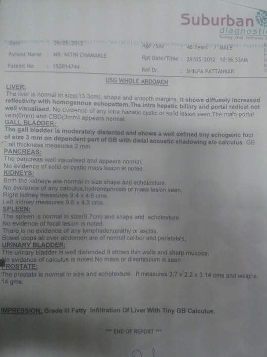
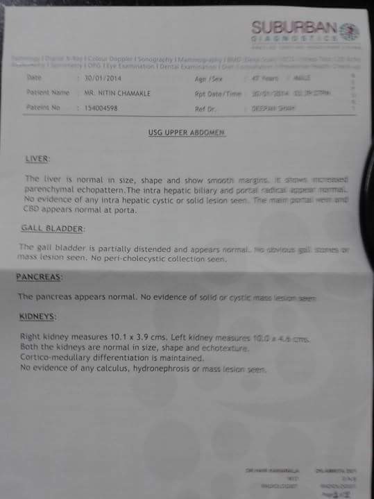
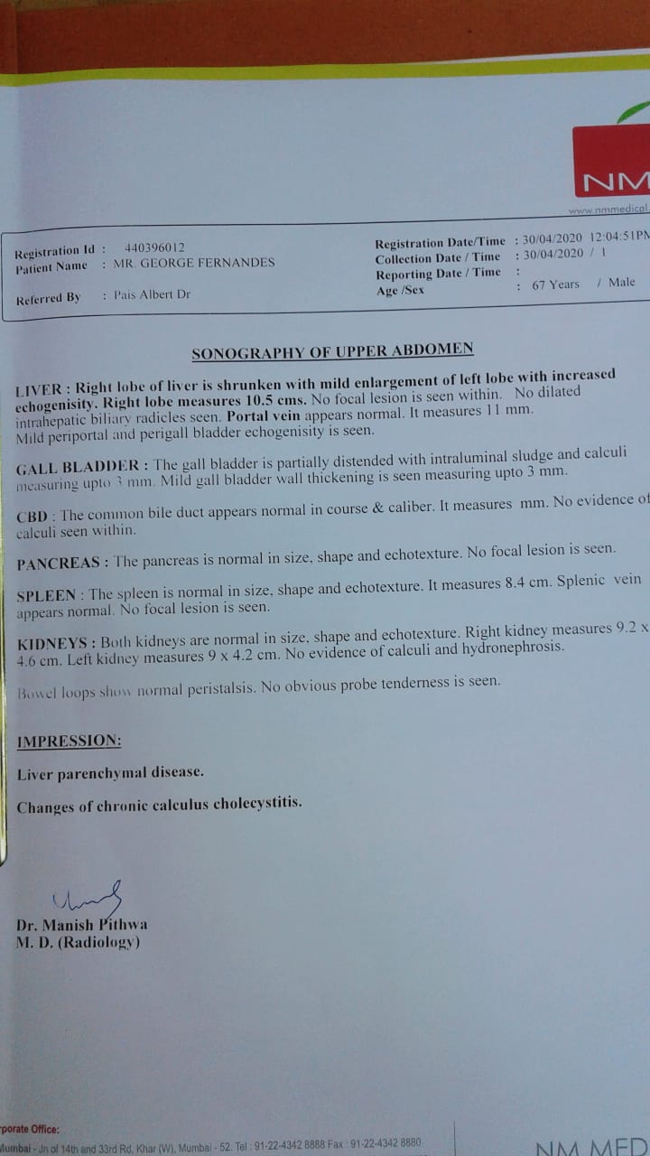
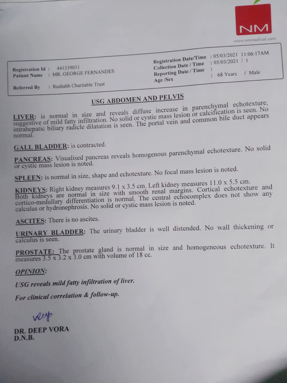
CASE A – GALL BLADDER STONE CURED WITH HOMEOPATHIC MEDICINES by Dr DEEPAN P SHAH -MD(Homoeopathy) at Dr SHAH’s Homeopathy -BEFORE TREATMENTCASE A – GALL BLADDER STONE CURED WITH HOMEOPATHIC MEDICINES by Dr DEEPAN P SHAH at Dr SHAH’s Homeopathy page-1 of reoprt AFTER TREATMENTGALL BLADDER STONE CURED WITH HOMEOPATHIC MEDICINES by Dr DEEPAN P SHAH at Dr SHAH’s Homeopathy page-2 of reoprt AFTER TREATMENTCASE B – Gall Bladder Stone USG report – Before treatmentCASE B – Gall Bladder Stone Cured With Homeopathic medicines USG report -After treatment
SYMPTOMS of GALL BLADDER STONES
Gall bladder Stones affect 10-20% of population in developed countries.
Upto 80% of Patients will never develop any symptom throughout their life and are called Silent Cases of Gall Bladder Stone and remainder 20 % become symptomatic at the rate of 1-3% per year.
Though most of the patient frequently present with non specific general mild complaints like eructations, GERD, unsatisfactory bowel clearance.
A few patients also present with prolonged mild discomfort in right hypochondrium and lumbar region with occasionall in epigastric region, with sensation as if weight or stone was lodged in the site of discomfort which aggravates on false stepping or jumping , unsatisfactory bowel clearance and other mild digestive derangements.
When Acute Symptom appear they are very severe and may be life threatening which needs immediate medical attention as in most of the cases it sets in whenever there are complications like
Cholecystitis- Inflamation of gall bladder due to infection or adhering of stone to bladder wall or severe statis and obstruction of downstream of bile due to blockage created by lodging of stone in bile tree, usually such blockages are created by very smallstones which can easily enter cystic and common bile ducts.
Inflamation of bile tree.
Pancreatitis is a very severe and life threatening condition may also present along secondary infection.
Gall Stone illeus when large stone enters adjoining loop of small intestine.
Empyema, collection of puss in pleural cavity due to secondary infections
Perforations
Fistulae
Patient with acute symptoms has severe excruciating spasmodic colicky pain in abdomen more pronounced in right hypochondriac region epigastric and umbilical region.
Fever with rigor and chills and it is indicative of secondary infection
DIAGNOSIS of GALL BLADDER STONES
Ultra Sonography is the most common prescribed test.
MRI in cases where unable to detect by USG or accidental finding.
CT Scan in cases where unable to detect by USG or accidental finding.
X rays not specific; very occasionally shadow is visualised in accidental findings, after which the patient is sent for USG for confirmation and proper evaluation of size and severity.
Serum Lipase and Serum Amylase activity with Ultra Sonography of pancreas need to be monitored frequently in cases with acute complaints to rule out pancreas involvement which can be life threatening.
MORPHOLOGY of GALL BLADDER STONE
Gall Bladder Stone consists 50-100% cholestrol.
More the cholestrol more pale yellow colour of stone, Exclusive Cholestrol stones are complete pale yellow in colour.
More the proportion of Calcarea Carbonate and Phosphate, more the stone has Greyish-white to black discoloration from pale yellow.
BEST HOMEOPATHIC MEDICINES for GALL BLADDER STONES
CALCAREA FLOURICA
Calcarea flourica is a powerful tissue remedy. Calcarea flourica is a combination of lime and flouric acid. It is a naturally occuring mineral in the body. Calcarea flourica is very useful remedy in the treatment of gallstones. Usually patient presents with prolonged mild discomfort in the right hypochondrium and lumbar region. Gastric derangements, weakness, loss of appetite. Vomiting of undigested food. Indigestion, fatigue and brain fag. Presents with paroxysms of lancinating paind in the right hypochondrium awaking him about midnight with aggravation lying on painful side. Sensation of dull weight and discomfort in the right hypochondrium. Sensitive to cold, to drafts and changes in weather.
FEL TAURI
Fel tauri is remedy used since ancient time. Fel tauri is a very efficient remedy for gallstones. Fel tauri increases the duodenal secretions, emulsifies fats and increases the peristaltic action of intestine. Liquifies bile and acts as a purgative. Presents with disordered digestion, diarrhoea, obstruction of bile ducts. Eructations and pain in the epigastrium. Gurgling in stomach and epigastric region. Pain in the right hypochondrium with sensation of heaviness. Fel tauri liquifies bile and helps in expulsion of biliary calculi.
CHOLESTRINUM
Choleatrinum is considered as a specific remedy for gallstones. It helps to relieve pain at once. Useful in obstinate hepativ engorgements, obstinate gall stones. Pain in the right hypochondriac region. Sensation of heavines in the abxomen. Gastric derangements. Reflux of gastric contents with pain in the epigatrium.
SILICEA
Silicea is a deep acting remedy indicated in case of gallstones. Silicea disturbs tbe stomach, causes hiccoughs. Severe pain in abdomen relieved by heat, distension of abdomen with flatulence and rumbling. Tightness accross the abdomen. Disturbed by pressure of clothes and worse after eating. Constipation from inability of rectum to expel faeces. Great straining at stool. Constant urge but cannot expel requiring mechanical aid. Hardness and distension of the region of liver with throbbing pain. Gastric derangements causes hicoughs, nausea, vomiting, disturbs the liver. Silicea is usually very sensitive to extremes of heat and cold.
CARBO VEGETABILIS
Carbo vegetabilis is a very effective remedy in patients suffering from gallstones. When Carbo vegetabilis is indicated the patient will be below par, the digestion will be slow and imperfect. Heaviness and fullness sensation. Sensation of weight in the abdomen. All gone sensation not relieved by eating, but after a few mouthfuls there is a sense of repletion. There is great distension of abdomen which is temporarily releieved by belching. Burning in the stomach extending to back and along the spine. Cramping pain forcing patient to bend double.
CARDUS MARIANUS
The action of Cardus marianus is centered on the liver, gall bladder, portal system causing apin, soreness. Pain in the region of liver. Swelling of gall bladder with painful tenderness.Sensation of fullness and soreness. Constipation with hard stool, difficult knotty, alternates with diarrhoea. Poor appetite. Nausea retching and vomiting sensation. Inflammation of gall bladder, pain in the upper abdomen in the region of gall bladder which is accompanied by nausea and vomiting.
CHELIDONIUM MAJUS
Chelidonium majus is one of the most indicated medicine for gallstone. Effective medicine for gall stones and jaundice when there is obstruction of bile ducts.Useful when gallstone colic is shooting, stabbing, tearing and lancinating type extending to back. Colic with constriction as by a string across abdomen. Most important symptom of Chelidonium is constant pain under the right shoulder blade. Suffers from hepatic or gastric symptoms. The tongue is coated thick and yellowish with imprints of teeth. Alternate diarrhoea and constipation. All symptoms are usually right sided.
CALCAREA CARBONICA
Calcarea carbonica is one of the indicated remedy in gallstone. Calcarea carbonica is usually suited to fair, fat, flabby constitution. Shooting pain around the liver with discomfort after wearing tight clothing. Sensitive to slightest pressure. Liver region painful when stooping. Presents with severe gall stone colic. Weak digestion. Milk disagrees. Feeling of tumefaction and fullness. Loss of appetite. No desire for food. Constipation. Frequent nausea ezpecially in morning. Cramps in stomach at night. Pains generally shooting or tensive, pressive type with swelling and induration of hepatic region.
CRATEGUS OXYACNTHA
Crategus oxyacantha is a excellent heart tonic and also useful in gallstone colic. Acts a a tonic for heart muscles that have becomw weakened due to reduced blood supply to heart as a result of deposits of cholestrol in arteries. Crategus is a very useful remedy in dissolving cholestrol deposits in arteries. Colic with severe pain in the hypochondrium with sensation of heaviness. Presents with difficulty in breathing with slight exertion. Dyspepsia and nervous prostration. Cold extremities.
COLOCYNTHIS
Colocynthis is indicated in case of choleliathiasis. Colocynth is a ideal remedy when gall stone pain is of a cutting shooting nature and better by bending double or by applying pressure. Worse by cold. Presents with pain in right hypochondriac region with sensation of fullness of abdomen. Colocynth is usually a acjte useful in gall stone colic. Neuralgias with sense of restlessness. Sharp shooting and lightening type of pain.
SLE or Systemic Lupus Erythematosus or sometime only called Lupus is a chronic systemic autoimmune condition with genetics, nutrition and environment playing a major role in its onset and evolution, its a condition which greatly reduces life expectancy and also causes complications in pregnancy. It falls under disabling disease conditions category though much less than10% cases develops disabilities.
TYPES of SLE
Acute Cutaneous Lupus
Sub-Acute Cutaneous Lupus Erthematosus
Chronic Cutaneous Lupus also called Discoid Lupus Erthematosus
Neonatal Lupus Erythematosus
Drug Induced Lupus Erythematosus
EPIDEMIOLOGY of SLE
Lupus was the most google searched topic in healthcare related topics in 2017! Now that is something to be added in Epidemiology section! 😀 hehehe!
Female are affected more with SLE than male, female of child bearing age are affected more with female to male ratio of about 9:1.
African, Caribean and chinese ethnic groups are more prone to this condition.
SIGNS and SYMPTOMS of SLE
GENERAL
Undifferentiating symptoms which are common to other diseases as well.
Fever
Fatigue
Malaise
Joint pain
Muscle pain
Bodyache
Sleep disturbances
Psychiatric Disorders
Poor physical fittness
Anaemia of chronic disease
Raynaud’s Phenomenon
SKIN
Majority of cases shows skin manifestation of the condition
Though the rashes and lesion on skin may vary, the classic sign of SLE on skin is butterfly rashes on face, also called Mallar Rashes and is seen in almost half of the cases with skin lesions.
If it is Acute type there might not be scaling of skin, not well demarcated distinct edge compared to othe types.
If it is Subacute type rashes shows scaling of skin with distinct edges.
And in Chronic type rashes shows thick distinct thick scaling with very well demarcated edges.
Increased Hairfall is also one of the complaints.
Ulcers in mucous membranes esp of nose and mouth.
MUSCULOSKELETAL
It affects Small joints especially of fingers and wrist and it very closely mimicks rheumatoid arthritis and psoriatic arthritis making it clinically difficult in making differential diagnosis.
But it is less destructive and disabling compared to other two conditions, only less than 10% cases of SLE will develop deformities and even fewer will have disabilities.
It not only mimicks Rheumatoid Arthritis but it also seems to have very close relation to rheumatoid arthritis.
It significantly increases risks of fractures and also it is associated with facilitation of Osteoarticular Tuberculosis.
HEAMOTOLOGICAL
Anaemia of chronic disease with low Red Blood cell count
Low White Blood Cell count
Anti phospholipid antibody syndrome is a condition where anti-phospholipid antibodies increases Partial Thromboplastin time causing tendency to heamorrhages and it is frequently found positive in patient with SLE and its coexisting is termed as Lupus Anticoagulant Positive.
Patients with SLE frequently show positive results for Anti Cardiolipin Antibodies as well, and patients with these antibodies sometimes shows false positive results for Syphillis.
CIRCULATORY SYSTEM AND HEART
Artheroscelerosis – Thickening and deposition of cholestrol plaque in blood-vessel walls which may also give rise to Ischemic Myocardial diseases like Myocardial Infarction.
Vasculitis – Inflamation of blood-vessels in some cases
Endocarditis – Inflamation of inner linning of heart, when due to SLE its termed as Libman-Sacks Endocarditis
Pericarditis – inflamation of outer layers and and surrounding tissues.
Myocarditis – Inflamation of cardiac muscles.
It may also cause inflamation of Mitral and Tricuspid valves of heart.
LUNGS
Pleurisy – Inflamation of pleura.
Pneumonitis – Inflamation of lungs.
Interstitial Lung Disease
Pulmonary Embolism
Pulmonary Heamorrhage
All these complications and chronic inflamatory processes causes Shrinking Lung Syndrome where there is reduction in lung volume.
REPRODUCTIVE
30 % of pregnancy has comolications like
Fetal Death
Spontaneous Abortion
Still Birth
Prognosis is worse in those who get aggravations in SLE duringbpregnancy.
Neonatal Lupus Erythematosus
Child born to mother with SLE shows symptoms of Discoid Lupus Erythematosus with
Heart block
Splenomegaly – Enlargement of Spleen
Hepatomegaly – Enlargement of Liver
Neonatal SLE is self limiting condition and in most cases recovers on its own.
RENAL (Kidney)
Painless Heamaturia – Blood in urine.
Painless Proteinuria – Protein in urine.
Lupus Nephritis leading to terminal Renal failure.
Histologically it shows its classical appearance of Membranous Glomerulonephritis with Wire-Loop Abnormailities due to deposition of Immune Complexes in Basement Membrane.
NEUROPSYCHIATRIC (NP-SLE)
If any Neuro-Psychiatric is caused dur to SLE its called NPSLE Neuro-Psychiatric Systemic Lupus Erythematosus
There are atleast 12 Central Nervous System related and 7 Peripheral Nervous System relate manifestation of SLE that are being observed in patients with SLE.
SLE is considered amongst one of the prototype disease as its very difficult to differentiate from many other autoimmune conditions as they share in common majority of signs and symptoms making its diagnosis very difficult, It much depends on clinical picture of the disease and investigations after that there are many criterias based on clinical symptoms coupled with laboratory tests based on which a person can arrive at some conclusion though not absolutely sensitive and specific to confirm diagnosis in every case but fair enough.
LABORATORY TESTS
ANA detection by direct or indirect immunoflorescence
ANA test detects many different subtypes of ANA related to many other autoimmune conditions as well with many overlapping eachother of which
Anti- Double Strand DNA Antibodies most specific of all present in almost 70% cases of SLE with only 0.5% non-SLE cases has t in them.
Anti-Smith DNA Antibodies present in most of the cases of SLE and not frequently found in non-SLE person.
Anti- Histone Antibodies present in Drug Induced SLE
Anti- U1 RNP antibodies – non specific also appears in other conditions like Systemic Sclerosis
Anti- Ro or SS-A and Anti- La or SS-B – non specific for SLE but more Specific to Sjogrene syndrome, but its present in many of the cases of neonatal lupus with heart involvement in particular.
Other Tests
Anti-ENA Test
Lupus Cell Test – It was used in past as it used to show positive in 50-70% SLE cases but was not specific to SLE and used to be present in many cases of many other conditions like RA Scleroderma etc.
DIAGNOSTIC CRITERIA OF AMERICAN COLLEGE OF RHEUMATOLOGY.
Its a stringent criteria developed by American College of Rheumatology, so that non of non-SLE cases should filter in, so many of the SLE cases are also filtered out.
The criteria is that if any patient shows any of the four symptoms out of eleven simultaneously or serially in more than one occasion than he is considered to be positive for SLE.
Mallar Rash/ Butterfly rash on cheeks; Sensitivity of 57% and Specificity of 96%.
Discoid Rash; Sensitivity of 18% and Specificity of 99%.
Serositis, Inflamation of serous membranes around heart (more specific) and lungs(more sensitive); Sensitivity of 56% and Specificity of 86%.
Mucosal Ulcers of oral cavity and nasopharynx; sensitivity of 27% and specificity of 96%.
Arthritis, non-erosive with more than two joints involved with tenderness swelling and effusion; Sensitivity of 86% and Specificity of 37%.
Photosensitivity, Ligh causes aggravation in skin rashes or other Lupus related complaints; Senitivity of 43%and Specificity of 96%.
Non Drug Induce : Hemolytic Anaemia, Leucopenia, Lymphopenia, Thrombocytopenia; Sensitivity of 59% and Specificity of 89%.
More than 0.5g of total protein in urine in a day or cellular cast seen in urine under microscope; Sensitivity 51% and Specificity of 94%.
Anti- Nuclear Antibody positive; Sensitivity of 99% and specificity of 49%.
Positive Anti- Smith, Anti- Double Strand DNA, Positive Anti- Phospholipid Antibody, False Fositive Serological test for Syphillis; Sensitivity 85% and Specificity of 93%; Presence of Anti- ssDNA in 70% of cases.
Neurological disorder Seizure or psychosis; Sensitivity of 20% and Specificity of 98%
This is a very stringent criteria used for research purpose if we go through we may falsely conclude negative diagnosis and miss out on diagnosing many patients who are suffering from SLE
Aslo it misses out on certain factors like antiphospholipid anti bodies which has strong association with SLE there are many cases who are anti phospholipid antibody positive but are not fitting in above ACR criteria but still they are having SLE.
So more practicle approach widely used is through the Recursive Partitioning which has two classification trees
The Simplest Classification Tree – If patient has any immunological disorder with positive anti- Smith antibody, anti- DNA antibody, false positive serology test for Syphillis, presence of Lupus cells or Mallar rash/butterfly rash, then the person is diagnosed as positive for SLE; specificity of 92% and sensitivity of 92%.
Full Classification Tree : It uses six criterias; sensitivity of 97% and specificity of 95%
HOMOEOPATHIC MEDICINES FOR SLE
I have seen homoeopathy work wonders in SLE especially in cases with NPSLE because then the disease becomes very expressive about itself, it shows itself not only on skin and joints but also on Neuropsychiatric sphere which show various symptoms typically different in each individual and this is what is required in homeopathic medicine selection, that the body is expressing itself in mental sphere which makes remedy selection easier.
Always a proper case taking needs to be done in deep seated chronic autoimmune conditions like this and a deep acting polycrest remedy should be selected after proper repertorisation as per each individual constitution and constitutional treatment is the only permanant solution for such conditions.
Still some theraputic indications are given which can be helpful guide and can be used as per the symptomatology in course of disease if indicated intercurrently or during acute excerbations of disease showing following symptom.
BELLADONNA
– Typically suited in Mallar rash or Bitterfly rash of Syetemic Lupus Erythemotosua with symptoms of Neuro-Psychiatric SLE (NPSLE) where CNS involvement is markedly noted also suits in PNS symptoms of NPSLC
MERCURIOUS SOLUBILIS
Whenever in case of lupus there are oral and/or naso-pharyngeal ulceration this remedy is very well suited
BORAX
Again this is best suited in mucosal ulceration but in this remedy the ulceration are more marked in oral mucosa than in nasopharynx.
SYPHYLLINUM
A nosode a dose can be given intercurrent as anti miasmatic of the cases that shows syphillitic miasma in the background also useful in cases showing painless red mallar rash or butterfly rash with much thickening and exfoliation especially like in Chronic Lupus Erythemotosus, also suited well in ulcerations of oral and nasopharyngeal mucosa.
CINCHONNA OFFICINALIS
In cases with signs of hemolytic anaemia wether due to disease ot allopathic medicines, it will work wonders in both the cases.
FERRUM PHOSPHORICUM
Where the patient has febrile condition due to disease with malaise, fatigue, Hairloss and aneamia duw to lupus or its medicines, can also be givem in low potencies in biochemic form along with other medicines
FERRUM METALLICUM
Red acute rash typically in acute lupus erythematosus with involvement of oral mucosa, also in later chronic stages when there is are signs of anaemia of chronic disease of hemolytic anaemia
RHUS TOXICODENDRON
Works wonders in cases of lupus where it not only acts on skin but also wonderfully acts on the joints and musculoskelwtal complaints the disease shows.
HYOCYAMUS
In patients with symptoms of NPSLE.
ACONITE NAPELLUS
in acute violent spells of relaopse and aggravations with symptoms of NPSLE like mental restlessness and has fear fright and anxiety in general and fear of death in particular.
Nipah Virus (NiV) Outbreak In India Claims 13 Lives
Nipah Virus infection outbreak,which is fast spreading and has fatality rate 0f 50-70%, has gripped Kerala (Kozhikode) in fear of wider epidemic.
Already 13 people are reported dead till now, of which 3 of them were from same family.
What Is Nipah Virus Infection?
Nipah Virus Infection is Viral Zoonosis that means infectious diseases that can be transmitted transmitted to Humans from Animals.
Nipah virus was first found in Pteropid Fruit Bats of Pteropodidae family.
Nipah virus is a RNA virus of Paramyxoviridae family of Order Mononegavirales and belongs to Genus- Henipavirus of species Nipah and virus name Nipah Virus (Niv).
In Humans Mortality Rate is very high , as per statistics of past outbreaks, almost 50-70% of patient infected will die.
The first documented outbreak of Nipah Virus Infection in human was in 1998 in Malaysia leaving 100 dead at that time.
Till date less than 600 cases of human infection of NiV are reported globally of which almost 60% had died.
Causes Of Nipah Virus Infection
Direct Contact with infected Humans or Animals, especially Fruit Bats and Pigs.
Half eaten fruits by Fruit Bats.
Contact with Fruit Bat droppings.
Contact with body secretion of infected Humans or Animals.
Signs and Symptoms of Nipah Virus Infection
Nipah Virus Infection in humans is very fast progressing fatal condition.
Incubation period of Nipah virus is 3-14 days during this period the first symptom starts to appear.
It presents itself in very initial phase as
Fever
Headache
Many cases presents breathing difficulty and other respiratory complaints these patients are more contagious compared to those who do not show respiratory complaints.
Few cases may have Pain in Abdomen, Nausea and vomitting.
Followed by symptoms of Central Nervous System (CNS) involvement, Encephalitis.
Drowsiness
Disorientation
Mental Confussion
In some cases blurred vision
And soon within 24-48 hrs of initial first symptom patient goes into Comma.
Diagnosis of Nipah Virus Infection
In case of an Epidemic or High Risk Group, it should be suspected in patients showing signs of illness and soon diagnosis should be established using Real Time Polymerase Chain Reaction (RT-PCR) of the sample taken from Throat Swab or Cerebrospinal Fluid, which directly detects virus proteins.
Viral RNA can be isolated from the saliva of infected person.
IgG and IgM antibody detection is possible only if the patient has survived the NiV Infection as most of the patients succumb to death even before this antibody appears in blood so its done in those patients who have survived and are on recovery phase and had not done RT-PCR for diagnosis, so just for confirming NiV Infection in those patients this antibody IgG and IgM tests are done.
And in those who have died and are suspected of death from NiV Infection, on those dead bodies, an Immunohistochemistry of tissue collected during autopsy is done this helps to get exact numbers of death due to disease and establish the scale of epidemic.
Treatment of Nipah Virus Infection
In Allopathy there are no medicines available against Nepah Virus and the treatment is based usually only on supportive measures.
In homoeopathy there are medicines available which are indicated in Nipah Virus Infection diesase condition.
Homoeopathic Medicines for Nipah Virus Infection
Very fast acting acute remedies, with profound effect on Central Nervous System and Respiratory system, needs to be selected for treating patients with nepah virus infection.
The best Homoeopathic remedies that are indicated as per Genus Epidemicus in Nipah Virus Infection Outbreak are
Notice :- Under mentioned Homoeopathic Medicines should be taken only under guidance and observation of qualified and registered homoeopathic practitioner.
Aconitum Napellus or Belladonna are suited best in initial stage initial 12-24 hrs of onset of first symptom when patient shows initial signs of illness that is fever and headache, this medicines can oslo be started in doubted cases without any side effects. (Note: Indian Homoeopathic Medical Association IHMA- Kerala based on its study report has proposed BELLADONNA to be probable PREVENTIVE HOMEOPATHIC MEDICINE FOR NIPAH VIRUS INFECTION in humans)
Gelsemium Sempervirens is best indicated when symptoms of central Central nervous system have developed like Dullness, Diziness, Drowziness, Disorientation, Blurring of vision tremors and trembling.
Nyctanthes Arbortristis in Homoeopathic Mother Tincture from is indicated in general.
Plumbum Metallicumor Ipecacuana is indicated in patient who shows symptoms of pain in abdomen, nausea, vomitting.
Aspidosperma Quebracho or Blatta Orientalis or Arsenicum Album are indicated homoeopathic remedies in patients showing symptoms of respiratory tract involvement with breathlessness and choking sensation.
Stramonium or Baptisia or Zincum Metallicumare best suited in later stages qhen patient is in delerium or comatose state.
Along with these homoeopathic medicines it is necesarry to isolate patient under hospital admission medical observation with proper supportive and symptomatic treatment.
Prevention of Nipah Virus Infection
Isolate infected persons.
Avoid travelling to affected regions.
Make sure food and drinks might not have been contaminated by bats.
Avoid eating raw fruits.
Do not eat fruits seems to be damaged by Fruit Bats.
Stay away from Bat and other animal droppings.
Do not drink Taddy brewed in open containers near palm trees.
Avoid Meat and Animal Products in the region of outbreak.
Maintain proper cleanliness and self hygiene especially sanitary hygiene.
Wear N95 mask when at public places if you are working, staying or travelling in region of outbreak.
Avoid travelling to regions having fruit bats, especially caves and under those trees where bats resides.
Psoriatic Arthritis is a condition found in patient suffering from autoimmune disease called Psoriasis where in there is involvement of joints as well, along with skin and nail. It is classified as Seronegative Arthropathy and individuals with HLA B27 genotype shows more susceptibility towards this condition with genetic and environmental factors playing a major role in precipitation of the condition.
It presents itself as Chronic Inflamatory Arthritis involving one or more joints which accompanies or alternates with acute of spells of Psoriasis or has relation with the cyclic pattern of Psoriasis.
Signs and Symptoms of Psoriatic Arthritis
If a person is suffering from Psoriasis doesnt mean he will develop psoriatic Arthritis, regardless of severity of psoriasis he is suffering from. But its observed that person having Psoriatic Arthritis shows some relation between skin complaints and joint complaints either they aggravate together or alternate each other in their spells of aggravation but not in every case and always.
70% of Psoriatic Arthritis cases presents first sign of disease as psoriasis lesion on skin, in majority cases it usually develop almost 10yrs after onset of appearance of skin psoriasis. majority of them are adults between 35-55yrs.
15% first develops joint complaints then skin psoriasis develops later. and majority of them are children.
15% presents both skin and joint complaints originating simultaneously at the onset of disease.
Silverish scaly skin exfoliating cyclically leaving behind clear red base on extensor surface of skin and typically on scalp, around umbilicus.
Exudation and cuts may also be seen in severe cases.
Onycholysis, Ridges on nail, Pitting on nail, Hyperkeratosis of skin below nails of fingers and toes.
MUSCULOSKELETAL
It more frequently involves small joints of hand and feets especially of Fingers and Toes frequently wrist and spine are also involved, There is morning stifness pain and swelling of joints. Almost 20% cases shows symetry in joints involved.
Swelling of fingers, Dactilitis, giving sausage like appearance to fingers.
Sacroilitis, Lumbar spondylitis, pain in lower back
Cervical Spondylitis, pain in cervical region.
Enthesitis of Tendo-Achilles causing severe pain in ankle.
Plantar Fascitis causing pain and stiffness in soles.
GENERAL
When the disease progresses or aggravates patient may show signs of
Malaise, Fatigue Weakness and Exhaustion.
Deformities, Disfigurement and Disabilities.
Anaemia of Chronic Disease.
Psoriatic Arthritis presents itself in one of the following five patterns.
1) Oligoarticular
Its the most common type affecting almost 70% of all the cases where in ther is involvement of less than 3 joints and lacks symetry.
2) Polyarticular
Its comparatively severe type and accounts for almost 25% of all the cases of which 50% develops disability and deformities. It typically involves more than 4-5 joints and shows somewhat symetrical pattern. It resembles much to Rheumatoid Arthritis and need to differentially diagnosed which becomes difficult in sero-negative Rheumatoid Arthritis cases.
3) Arthritis Mutilans
Also called Chronic Absorptive arthritis ans is found in almost 5% of all the cases and is the most severe and destructive form of arthritis presented due to psoriasis and in many other conditions like Rheumatoid Arthritis and it shows severe disfigurement and deformities.
4) Spondyloarthritis
Typically affects spine especially the cervical spine and sacro-illiac joint. may also affect other joints as well in a symetric fashion.
5) Distal Interphalangeal
Typically involves distal small joints of fingers and toes with involvement of nails.
Diagnosis of Psoriatic Arthritis
There is no Specific Test Available For Psoriatic Arthritis. It cant be predicited based either only on joint or only on skin complaints that the person is developing Psoriatic Arthritis and its only after both skin and musculoskeletal complaints presents itself the diagnosis can be established. So, early diagnosis is not possible and its usually established only after the disease has progressed enough to show itself on both the spectrums.
The diagnosis of Psoriatic Arthritis depends upon clinical features and host of investigative tests that collectively exclude probability of other conditions and indicate towards Psoriatic Arthritis.
Psoriasis with chronic inflamatory arthritis of which the aggravation pattern can be related to cyclic process of psoriasis but not necessarily in all cases. Especially showing features like
Distal interphalangeal arthritis
80% of patients presents nail complaints Onycholysis – ridging pitting and hyperkerotosis of skin under nail
Dactilitis, sausage like appearance of fingers.
30-50% patients show Enthesitis, involving Tendo-Achilles, Plantar fascia,
Pain around Patella, illiac crest, epicondyles, supraspinatus insertions, sacroillitis.
X ray shows degenerative changes
Family history of psoriasis or psoriatic arthritis
As mentioned above its an autoimmune condition chronic in nature. So its important to treat the cause first and the symptoms will be relieved on its own. For that a proper homoeopathic case taking and constitutional approach is necessary as its deep seated genetic complaint and only a deep seated constitutional remedial force can bring about change for better. But in many severe cases where there are gross degenerative changes or the disease has progressed much further or the pain is severe we need to treat theraputically initially to get the acute exacerbations in check and later we can find out constitutional remedy based on Miasmatic background and totality of symptoms of Mind and Body. Also There arise much need for anti-miasmatic nosode administeration in the course of treatment if the case is improving but improvement is soon regressing or case has hit a stand still and ia not reaponding further in such case we need to consider a nosode.
Homoeopathic Medicines for Psoriatic Arthritis
RHUS TOX
Rhus tox is usually adapted or well suited to a person of rheumatic diathesis. Rhus tox is very well indicated in psoriatic arthritis. The effects on the skin, rheumatic pains, mucus memmbrane affections makes this remedy frequently indicated. Rhus tox affects the fibrous tissue of the joints, ligaments causing rheumatic symptoms. Patient presents with burning eczematous erruptions with tendency to scale formation. Skin is red swollen with intense itching. Hot painfull swelling of joints. Rheumatic pains spread over a large surface area at nape of neck, loins, and extremities which is relieved by motion. The cold fresh air is intolerable makes the skin painful.
URTICA URENS
Urtica urens is one of the indicated remedy for psoriatic arthritis. Rheumatism is usually associated with skin complaints. As its common name stinging nettle implies it produces stinging and burning pain. Skin complaints alternate with rheumatism with severe pain in joints. Itching and swelling all over body resembling hives. Heat in skin of face arms shoulderand chest with formication numbness and itching. Psoriatic errruptions with scales, skin looks wrinkled. Intense burning of skin. Patient is worse from exposure to cold moist air.
RHUS VENETA
In case of Rhus veneta, the skin symptoms of this species of rhus are most severe. Rhus veneta is one of the most actively poisonous remedy among the family. Presents with great restlessness.,numbness and lameness of joints. Bruised feeling in the limbs. Pains as if sprained or dislocated. Presents with trembling of limbs with twitching of muscles. Rashes under the skin with severe nightly itching. Fine psoriatic erruptions on forearm, wrist, back of hands between and on fingers. Severe desquamation with severe itching. Complaints are usually aggravated by warmth.
LEDUM PAL
Ledum pal usually affects the fibrous tissue of joints especially small joints. Hence it can be called as a rheumatic remedy where rheumatism begins in the feet and travels upwards. Ledum pal is very valuable remedy in psoriatic arthritis. There is weakness and numbness of affected parts. Painful cold oedematous joints. Presents with erruptions only on the covered parts of the body. Affecting the skin ledum produces reddish spots with scaly erruptions. Gouty pains shoot all thrkugh the foot and limbs. Cracking in joints worse from warmth of bed. Aggravation from warmth is so severe that the patient can only get relief from rheumatism is by putting his feet in cold water.
BELLADONA
Belladona has marked sphere of action on the skin, bones, glands and nervous system. Useful in case of psoriatic arthritis. Pains are usually throbbing, sharp, cutting, shooting which come and go in repeated attacks. Joints are swollen, red hot with severe throbbing sensation and extreme sensitiveness. The heat, redness and burning characterise most of the skin complaints, and presnts with alternate redness and paleness of skin with scaly erruptions and severe itching. The complaints of belladona come on suddenly, eun a regular course and subside suddenly.
SILICEA
Silicea too can be considered one of the efficient remedy in psoriatic arthritis. Silicea produces inflammation of skin. It acts upon the constitution that are sluggish. There is i.perfect assimilation and defective nutrition. Presents with neurasthenic states and increased susceptibility to nervous stimuli. Presents with moist erruptiions on skin with fomation of scales. Usually patient presents with weak spine, susceptible to draught on back. Pain in coccyx. Diseases of bones of spine.
MEDORRHINUM
Medorrhinum is a nosode prepared from gonorrhoeal virus. It is a powerful deep acting remedy indicated for most of the cgronic complaints. Medorrhinum is a very valuable remedy for arthritic and rheumatic pains , loss of power in joints, joints feel loose. Useful in chronic psoriatic arthritis wuth great disturbance and irritabilty of nervous system. Pain in back with burning heat. Legs feel heavy and ache all night. Acting on skin medorrhinum causes intense itching worse at night. Yellowish copper coloured spots remain after erruptions.
ACTEA SPICATA
Actea spicata is a rheumatic remedy especially affecting the smaller joints, tearing tingling pain. Presents with wrist rheumatism. Though wrist is affected prominently other joints are too equally affected. Slight fatigue causes swelling of joints. Psoriatic erruptions on skin which are dry scaly and with intense itching.
SYPHILLINUM
Syphilinum is another nosode prepared from syphilitic virus. Acts on the bone, nerves, mucus membrane. Indicated in psoriatic arthritis. Presents with rheumatic stiffness and lameness in back. Aching in the whole spine. Inflammation of joints. Pains are usually aggravated by warmth of bed. Indiacted in shifting rheumatic pains and chronic eruptions like psoriasis, presents with dry scaly or pustular erruptions on different parts of the body in patches. Presents with great weakness with very few symptoms, utter prostration and debility in morning or on walking.
THUJA OCCIDENTALIS
Thuja has main action on the skin, bones. Useful in case of psoriatic arthritis. Thuja chiefly acts on the mucus membrane of skin, nerves, glands. Presents with cracking in joints when stretching them. Limbs feel as uf made of wood or glass and would break easily. Psoriatic erruptions itch or burn violently. Erruptions on covered parts of the body. Worse from scratching. Dry skin with brownish spots and scales with severe itching.
We will make a big mistake in understanding and treating the person if we ignore how all the subtle impressions can influence our body.
By the time you complete reading this article you will have a major change in your philosophy of perceiving your body and you will start having a more broader view and acceptance towards many other aspects of nature and its implications on health and Life.
Senses like Vision, Hearing, Smell, Touch, Taste generates feelings which generates desires and thoughts. Can thought generate itself in absence of all these senses?
Thought gives rise to emotions like Love, Hatered, Anger, Fear, Sadness, Happiness etc.
Many neurotransmitters like acetylecholine, Dopamine, Serotonine, GABA(Gama Amino Butric Acid), Glutamate, Norepinephrine and there are 60 more such neurotransmitters discovered by scientists who’s secretion is triggered by thought and vice-a-versa.
Senses, Thoughts and Emotions triggers neurotrasnsmitters that determines functioning of our brain which has hypothalamus and a pitutary glands adjoined below which is called master of orchestra of hormonal interplay in the body as it determines the level of each hormone in the body that we call Hypothalamo-pitutary-Hormonal axis. And you all know how important the hormones are seeing their profound effect on body’s functioning in general. Slightest disturbance in Hypothalamo-Pitutary-Hormonal axis results into grave, acute and chronic complaints.
As we all know that the body parts or organs that are being used more, develops better and those which are not used goes into disuse atrophy and later becomes dormant. Similarly certain functions of body change as per their necessity. We know that most of the body processes are triggered directly or indirectly by brain and our thoughts and emotions.
Certain examples of physiological impact of senses, thoughts, feelings, emotions and other abstract dynamic influences.
Sadness causes weeping, it activates tear glands.
Fear causes activation of sweat glands , shivering, piloerections, palpitations
Certain thought Sound and Vision causes stimilation of sexual organs
Stimulation of salivary glands on sight of food
Nausea on certain sight and odour
Depression causes severe appetite and eating disorders, Hypothalamus directly controls our hunger and satiety centers slightest disturbance in this physical psychological or other may cause eating disorders leading to obesity and or alarming weightloss.
Depression is thought to be one of the cause of hypothyroidism and vice-a-versa.
Now after years of reasearch and experience modern science has accepted the fact that most of the autoimmune conditions are triggered or caused due to psychological stress as one of the major reason.
All these examples shows us that how abstract dynamic influences external or internal has impact on brain and subsequently on our body’s physiology, the prolonged changes in physiology has a wider implication on body’s each and every cells and thus changing the gene environment and epigenome thus influencing gene and finally its structure.
We know the processes of adaptation, our body gradually adapts itself to the type of stimulus we are subjected to for a longer period of time. Similarly our thoughts emotions and other psychological factors influences our body’s physiology and its profound effect for a longer period acts as an effect of a stressor and body has to adapt itself accordingly. Now adaptation initially seemingly to be phenotypic may gradually change genotype as well. Now a change at one locus in gene will have compensatory and/or counter regulatory change at other locus as the harmony is spoiled and body tries to re-harmonize or may be due to one bad sector in gene damaging other sectors(locus) due to their abnormal protein products in gene itself factoring in new changes. Now this leads to accumulation of many changes in the character of genome giving rise to positive or negative or mixed physiological or anatomical variations or disturbances leading to diseases.
Majority of endogenous diseases are caused due to genetic imbalance that we got it as hereditary germ line change or are acquired by external impressions and influences that has profound impact on our body.