Category Archives: HOMEOPATHIC TREATMENT

Diseases and their homeopathic treatment.

FROZEN SHOULDER HOMEOPATHIC TREATMENT

Frozen Shoulder also called Adhesive Capsulitis of shoulder joint is painful stiffness limiting range of motion of shoulder joint caused due to inflammation related pathophysiology in capsule of shoulder joint.

CAUSES AND RISK FACTORS OF FROZEN SHOULDER

CAUSE OF FROZEN SHOULDER

Exact cause of Frozen Shoulder remains unclear but there are many factors that increases the risk and lead to frozen shoulder

RISK FACTORS FOR FROZEN SHOULDER

GENDER

Frozen shoulder is more common in females than in males

AGE

Person above 40yrs of age are more prone to this condition

INJURIES

  • Injury to shoulder joint involving any of its companent especially gleno-humeral joint capsule or rotator cuff.
  • Sports persons typically develop calcifications of tendons around shoulder joint and also in joint structures due to repeated injury and result of prolonged inflamation such persons are at high risk of developing frozen shoulder.

REDUCED MOBILITY

  • Injuries to arm and neck may indirectly affect shoulder joint, due lack of motion in general of that region as a whole and changes pattern of gait and movements of that region.
  • Immobility or reduced mobility post surgerr with general debility and delayed recovery.
  • Immobility post stroke.

MUSCULO-SKELETAL DEFECTS

  • Certain congenital structural anomaly of Musculoskeletal framework which has influence on movement, gait and weight distribution pattern on shoulder joint in abnormal pattern, which may put the person at risk of develooing Frozen Shoulder.
  • Kyphosis or Scoliosis or other congenital or acquired spinal anomaly especially those above thoracic spinal level may increase risk of frozen shoulder.
  • Patients with cervical spondylosis are at risk of developing frozen shoulder.

RHUEMATIC DISORDERS

OTHER SYSTEMIC DISEASES

SYMPTOMS OF FROZEN SHOULDER

  • Pain in affected shoulder aggravates on movement
  • Loss of rang in motion of affected shoulder joint

Frozen Shoulder is divided into 3 stages

1) Freezing Stage

In Freezing Stage there is pain in joint which aggravates on motion and he range of motion gradually starts decreasing.

2) Frozen Stage

In Frozen Stage the pain is not much as compared to freezing stage but the range of motion is completely diminished so much so that patient can barely move the joint or cant move at all.

3) Thawing Stage

In Thawing Stage pain subsides and range of motion gradually starts improving and complete recovery is established in few weeks.

If it is acted upon quickly in freezing stage with intervention of physiotherapy and proper medication the progress of the disease is arrested and soon resolves completely.

DIAGNOSIS OF FROZEN SHOULDER

On clinical presentation of symptoms of the patient frozen shoulder can be diagnosed and radiological tests like X-rays, CT scan and MRI may be suggested inrequired cases to find out extent, severity of pathological condition within and also to rule out any other pathological condition within or around shoulder joint.

If along with shoulder joint if there are other joints involved simultaneously or frequently different joints in past then it becomes necessary to rule out other underlying systemic conditions like Rheumatoid ArthritisGout, Ankylosing Spondylosis, Systemic Lupus Erythematosus etc and for that your doctor may suggest blood tests like RA factor, ESR, CRP, ANA, S.uric acid and HLA B27 (where there is severe spinal involvement along with shoulder). Vitamin D should also be checked wether its in optimal range for better recovery.

TREATMENT OF FROZEN SHOULDER

Treatment of frozen shoulder is based on combination of both Physiotherapy and Medicines.

Physiotherapy plays a major role in cases of frozen shoulder. Physiotherapist takes support of certain modalities, recomends certain exercises and manual physical intervention where in he may exert mild force or pressure along and within the axis of movement of shoulder joint and try to open up the freezed shoulder and gradually retain its laxity and mobility.

Pain may temporarily increase during and  after physical intenvention but with regular recomended exercise and medicines it finally subsides and joint regains its complete range of axis of movement subsequently.

HOMEOPATHIC MEDICINES FOR FROZEN SHOULDER

If the frozen shoulder is without any underlying condition specific medicines will work well but if it is secondary to some underlying condition like autoimmune diseases or metabolic disorders or Infections or some other systemic or acute or chronic complaints then constitutional trearment along with initial acute specific remedy may be required.

List of Common Homeopathic Remedies Used in Frozen shoulder

  • Calcarea Flourica
  • Silicea
  • Calcarea Phosphorica
  • Thiosinaminum
  • Rhus Toxicodendron
  • Bryonia Alba
  • Arnica Montana
  • Plantago Major
  • Belladonna
  • Ledum Palusture

LUMBAR SPONDYLOSIS

Degenerative changes in vertebral bodies and intervertebral disc at the level of L1 to S1 in vertebral cloumn affecting lumbar region of spine is called Lumbar Spondylosis.

It is characterised by degenerative changes of Lumbar vertebral bodies like osteophyte lippings and spur formation, degeneration of  intervertebral disc, narrowing of intervertebral space, Intervertebral disc bulging, intervertebral disc herniation.

It can also be called Osteoarthritis of Lumbar Spine.

CAUSES OF LUMBAR SPONDYLOSIS

Lumbar Spondylosis may be caused due to many factors that can be broadly classified into Mechanical, Congenital or Acquired Anatomic anomalies, Metabolic, Immunologic, Genetic, Nutritional, Ageing and Septic reasons.

Most common of all are ageing and mechanical reasons like excessive wear and tear due to wrong postural habbits, type of work like driving for long duration regularly, sitting for long hours, lifting weights or working with bent posture etc. that exerts much pressure on lumbar spine on regular basis so as to not giving much time to recover again and resulting into accumulation of wear and tear finally damaging the spine and causing lumbar spondylosis.

Metabolic problems like Hyperuricemia/Gout where high level of uric acid in blood damages lumbar spine also this uric acid may get crystalised to form monosodium urate monohydrate and settle in the intervertebral joints and damage them.

Immunologically mediated chronic inflamation in lumbar spine like in Rheumatoid Arthritis, Chickenguniya, Systemic Lupus Erythematosus etc.

Genetic Complaints like Ankylosing Spondylosis etc

Vitamin D deficiency causes reduced calcium assimilation resulting into reduced bone mineral density causing weak bones easy to damage. Insufficient nutrition compared to requirement, which prevents repair work of regular wear and tear of lumbar spine, due to which damages tend to get accumulated resulting into lumbar spondylosis.

Congenital or acquired Anatomical anomalies in musculoskeletal framework that may cause abnormal posture or abnormal weight distribution which results into pressure on spine in wrong fashion gradually facilitates development of Lumbar spondylosis.

Septic Conditions of spine like Tuberculosis of spine may result into lumbar spondylosis due to tissue lysis as result of infection.

SIGNS AND SYMPTOMS OF LUMBAR SPONDYLOSIS

  • Pain and Stiffness  in lower back or Lumbar Region.
  • Sciatica due to compression of nerve roots of sciatic nerve.
  • Pain stinging pain in lumbar spine
  • Pain radiating from lumbar spine to lower limbs
  • Paraesthesia in lower back genitals or lower limbs
  • Cramps calfs, thighs and other muscle and muscle groups of lower limbs
  • Difficulty in walking and maintaining posture and balance.
  • Lack of control on urinary spincter or anal spincter resulting into either retention of urine or bowel or involuntary expulsion of urine and bowel
  • Sexual debility Tabes Dorsalis

COMPLICATIONS OF LUMBAR SPONDYLOSIS

Myelopathy – When there is damage to spinal cord it causes Myelopathy.

Raduclopathy – compression of nerve near nerve root where it emerges out of spinal cord.

Vertebrobasilar insufficiency – Vertebral artery insufficiency due to its occulusion while it passes through vertebral foremen sue to degenerative changes for lumbar spine causes vertebrobasilar insufficiency leading to death of condrocytes of intervertebral disc and osteophytes starts accumulating and depositing.

DIAGNOSIS OF LUMBAR SPONDYLOSIS

  • Clinical symptoms if indicate towards Lumbar spondylosis it can be then confirmed with X ray.
  • MRI and CT scan gives us better imaging which helps  judge and evaluate the extent of damage and condition
  • Myelograph is dye injected while Xray and CTscan this gives detailed shadow to understand even better
  • Electromyography helps evaluate nerve involvement in spondylosis.

HOMEOPATHIC MEDICINES FOR LUMBAR SPONDYLOSIS

  • HYPERICUM PERFOLIATUM
  • CALCAREA CARBONICA
  • GNAPHALLIUM
  • CALCAREA PHOSPHORICA
  • MAGNESIA PHOSPHORICA
  • ARNICA MONTANA
  • NATRUM SULPHURICUM
  • RHUS TOXICODENDRON
  • SILICEA
  • MEDHORRINUM

CERVICAL SPONDYLOSIS

Our spine is classified into cervical, thoracic, lumbar and sacral regions.

Cervical Spondylosis is condition where in vertebra and intervertebral discs of cervical region undergoes degeneration. 

It is characterised by thinning of intervertebral discs, reduced intervertebral space, osteophyte lipping, spurs formations,  herniation of intervertebral disc, nerve compression etc.

It can also be called osteoarthritis of cervical spine.

CAUSES OF CERVICAL SPONDYLOSIS

This degeneration can be due to various mechanical, immunological, infective, metabolic, genetic, nutritional and age related reasons.

It can be due to one or combination of than one ot the above reasons. Most of the cases are due to ageing and mechanical wear and tear related to abnormal physical exertion and postural habbits.

It is commonly seen in people assuming wrong posture for long hours like lying down with head placed on huge pillow or watching mobile phones or reading books with tilted head for long hours, staying on computer workstation with an arm stretched on mouse or key board for long transfers weight on neck.

When such postural habbits are prolonged for few hours to days its starts inflamatory process in cervival spine and if still prolonged for months to years the prolonged inflamation and mechanical wear and tear results into degeneration of spine.

Many genetic and immune mediated conditions like Rheumatoid arthritis, Ankylosing spondylosis, Systemic Lupus Erythematosus, Psoriatic Arthritis etc may result into prolonged chronic inflamation of spine ingeneral and gradual degeneration of cervical spine as well resulting into cervical spondylosis.

Metabolic reasons like hyperuricemia may result into deposition of monosodium urate monohydrate crystals into joint spaces in cervical spine resulting into subsequent erosion and degeneration of spine causing cervical spndylosis.

Certain infections in spine like tuberculosis results into degeneration of spine and spondylosis and when it affects cervical region on spine it causes cervical spondylosis.

With ageing there is depletion of anabolic hormones and other factors required for quick repair process which results into slow repair process compared to daily wear and tear and damages tends to accumulate and gradually resulting into erosion and degeneration of spine.

Nutritional deficiencies arised due to lower intake compared to requirement, resulting into lower calcium vitamin D and many other nutrients which not only slows down the repair process to built up damages but also gives rise to low bone mineral density resulting into erosion and degeneration of vertebral bodies.

SIGNS AND SYMPTOMS OF CERVICAL SPONDYLOSIS

In initial stages it starts with occasional stiffness and pain in neck lasting few minutes to hours after exertion gradually it starts persisting with pain on extreme range of axis of movement of neck then later even on smaller axis or range of motion of neck patient starts feeling stiffness or pain or discomfort in neck

If not taken care the nerves originating from cervical plexus which emerge out from cervical spine they start getting compressed causing myalgia paraesthesia in neck which may radiate to shoulder and extend upto arm and upto tip of fingers.

In severe cases of cervical spondylosis patient may also experience vertigo nausea vomitting complete loss of balance with pain in neck and gastric derangement as concomittant symptoms

COMPLICATIONS OF CERVICAL SPONDYLOSIS

CERVICAL SPONDYLOTIC MYELOPATHY (CSM) – It is caused due injury to spinal cord due to cervical spondylosis.

CERVICAL SPONDYLOTIC RADICULOPATHY – In this the nerve gets pinched and compressed near the nerve root shortly after it leaves spinal cord.

VERTIBROBASILAR INSUFFICIENCY – When vertebral artery which is passing through vertebral formen gets occluded and deprives chondrocytes of intervertebral disc from circulation as a result the die and weaken intervertebral disc and osteophytes starts settling in.

DIAGNOSIS OF CERVICAL SPONDYLOSIS

Cervical Compression Test – When the neck is tilted laterally and applied downward pressure patient feels pain on ipsilateral side in neck or shoulder , its not conclusive but indicative and qualifies case for further radiological investigations

Lhermitte’s Sign – Electrict Shock like pain on flexion of neck

These patients general show reduced range of motion of n

Based on clinical symptoms patient may be sent for X ray  for ascertaining the diagnosis.

MRI and CT Scan helps to further find out extent and severity of damage to spine.

Myelography is done with dye injection in spinal cord during CT or X ray for more detailed radiological imaging

Electromyography and Nerve Conduction Test helps to find out involvement of nerve and nerve damage and extent of nerve injury.

TREATMENT FOR CERVICAL SPONDYLOSIS

Guidlines to patients on maintaining correct posture, avoid jerk and strain to secure neck is of utmost importance in management of cervical spondylosis patients.

Proper calcium intake sufficient exposure to sunlight for vitamin D. Increased protein, vitamin B12 and iron intake.

Exercise like pranayam and walk helps stimulate hormone secretion and thus facilitating absorption and assimilation of nutrients required for repair and rebuilding the worn and damaged tissues and to increase bone mineral density.

Mild gentle exercise of neck helps increase local blood flow and keep tissue supple and stimulate its growth and strenght but if not done under proper guidance of qualified physiotherapist it may further injure the already damaged tissue. So if proper qualified physiotherapist is not available to guide its safer bet not to exercise neck region involving cervical spine on your own and giving it complete rest and let it recover on its own while still continueing with walk and pranayam regularly.

HOMEOPATHIC MEDICINES FOR CERVICAL SPONDYLOSIS

CIMICIFUGA RACEMOSA / ACTEA RACEMOSA

RHUS TOXICODENDRONE

ARNICA MONTANA

PLANTAGO MAJOR

HYPERICUM PERFOLIATUM

TUBERCULINUM

BELLADONNA

CALCAREA PHOSPHORICA

BELLIS PERENIS

SILICEA

MEDHORRINUM

SWINE FLU H1N1

Swine flu is an infection caused by swine flu influenza virus which is common throughout the population worldwide. The disease affects the respiratory tract of pigs and is transmitted to humans.

THE SWINE FLU VIRUS

The influenza virus is a negative sense RNA virus of the family Orthomyxoviridae with three genera Influenza A, B, C.

Swine influenza is known to be caused by Influenza A subtypes. H1N1, H1N2, H3N1 H3N2. In pigs Influenza A virus are divided into subtypes based on 2proteins on surface of virus the haemagglutinin(H) and the neuraminidase (N). There are various types of subtypes of H and N.
Every year new strains of virus evade host defenses.
Another characteristic of type A influenza is that the virus has segmented genome with eight single stranded RNA segments. When the host cell is infected with more than one influenza virus, the genes have the opportunity to get ressorted and produce a different strain altogether. The antigenic shift is responsible for pandemic of influenza which has been observed in the past. Virus acquires the ability to not only infect but spread within the human host.

Influenza B viruses are only known to infect human and seals causing influenza

Influenza C virus infect both human and pigs and does not infect birds.

HISTORY OF SWINE FLU

Swine origin influenza A H1N1 virus was first recognised in the border area of Mexico and United states in April 2009 and during the span of 2 months and was spread with travellers wordlwide resulting in 1st influenza pandemic since 1968. Around mid 20th century identification of influenza sub types became possible allowing accurate diagnosis of transmission to humans. In 2010 the World Health Organisation declared Swine flu pandemic officially.
The term pandemic means that an infection has spread to many countries around the world causing widespread human suffering. This pandemic has become a worldwide concern. A new strain of Swine flu was detected in 2011, it did not affect a large number of people. In 2015-2016 a large outbreak of swine flu killed a huge number of people. Another outbreak 2017 affecting various people at different places and most recent is 2018 outbreak

The influenza virus is a negative sense RNA virus of the family Orthomyxoviridae with three genera Influenza A, B, C.

Swine influenza is known to be caused by Influenza A subtypes. H1N1, H1N2, H3N1 H3N2. In pigs Influenza A virus are divided into subtypes based on 2proteins on surface of virus the haemagglutinin(H) and the neuraminidase (N). There are various types of subtypes of H and N.
Every year new strains of virus evade host defenses.
Another characteristic of type A influenza is that the virus has segmented genome with eight single stranded RNA segments. When the host cell is infected with more than one influenza virus, the genes have the opportunity to get ressorted and produce a different strain altogether. The antigenic shift is responsible for pandemic of influenza which has been observed in the past. Virus acquires the ability to not only infect but spread within the human host.

Influenza B viruses are only known to infect human and seals causing influenza

Influenza C virus infect both human and pigs and does not infect birds.

EPIDEMOLOGY AND HISTORY OF SWINE FLU VIRUS INFECTION OUTBREAKS

HISTORY

Swine origin influenza A H1N1 virus was first recognised in the border area of Mexico and United states in April 2009 and during the span of 2 months and was spread with travellers wordlwide resulting in 1st influenza pandemic since 1968. Around mid 20th century identification of influenza sub types became possible allowing accurate diagnosis of transmission to humans. In 2010 the World Health Organisation declared Swine flu pandemic officially.
The term pandemic means that an infection has spread to many countries around the world causing widespread human suffering. This pandemic has become a worldwide concern. A new strain of Swine flu was detected in 2011, it did not affect a large number of people. In 2015-2016 a large outbreak of swine flu killed a huge number of people. Another outbreak 2017 affecting various people at different places and most recent is 2018 outbreak.

Influenza A outbreaks occur almost every year although their extent and severity vary.
In the last century, influenza virus caused 3 pandemics, the 1918 spanish flu, 1957 Asian flu, 1968 Hong kong flu. The out breaks differ in extent of spread.
The H1N1 strain responsible for current outbreak of swine origin influenza was first recognised at Mexico and US in 2009, and during a very short spam Swine flu became the 1st pandemic of 21st century. The outbreak has been pandemic in true sense involving more than 170 countries spread all over continents.
As per WHO there is a high risk of overall transmission of Swine flu globally.

SWINE FLU TRANSMISSION

As Swine flu is a viral disease it usully spreads from person to person by inhalation or ingestion of droplets containing virus from affected people who sneeze or cough leading to contamination of air with millions of virus in air.

CLINICAL REPRESENTATION OF SWINE FLU.

Signs and symptoms are similar to seasonal influenza virus or similar to other viral respiratory illness.
A common element of swine flu is upper respiratory symptoms. The patient presents with severe cold, stuffy or running nose combined with head congestion along with watery eyes associated with itching. Sore throat.
Severe bodyache, includes muscle weakness. Feeling of general malaise or weakness are very common with overall discomfort. The patient usually feels so weak that it hampers his daily activities. Presents with continous drowsiness.
In some cases of swine flu, gastrointestinal symptoms like stomach ache, cramps, nausea, vomiting and diarrhoea occur.

PREVENTION

  • Patients affected with swine flu should take necesary precautions.
  • Stay at home if you are sick.
  • Maintain distance from people so that you do not pass the infection to others.
  • Always wear a well fitted masks in public places.
  • Cover your mouth and nose while coughing and sneezing.
  • Wash your hands with soap. Make sure you wash your hand for atleast 15 sec. Use an alcohol based gels or foam sanitizers.
  • Use disposable tissues
  • Take proper rest.
  • Necesary preventive measures in people who are travelling or residing in areas where a swine flu outbreak have occured
  • Avoid close contact with people who have been affected with swine flu.
  • Always cover your mouth while coughing or sneezing.
  • Wash hands frequently as chances of infection through contact.
  • Maintiain proper hygeine.
  • Practice a healthy lifestyle habits such as nutritious diet, proper sleep, excercise.
  • Drink plenty of fluids.

DIAGNOSIS OF SWINE FLU

Swine flu is diagnosed based on the clinical representation of patient. Diagnosis can be made if the person has recently travelled to an area where there is Swine flu outbreak in humans.
If you residing in locality where there is epidemic of Swine flu, or if you come in contact with the influenza virus and symptoms of flu appears.
First thing you need to do is Rapid Influenza Diagnostic Test.. It is a blood test which detects whether the flu is of type A or type B. If type A turns to be positive then you should further go for PCR test for confirmation of subtype. It is advisable to straight away go with PCR in high risk group and in epidemic prone areas.

INSOMNIA – Sleep Management Schedule – Get Rid Of Sleeping Pills

Sleep Management schedule is designed to help insomnia patients who have difficulty in falling asleep or difficulty in maintaining state of sleep. It helps to avoid sleeping pills and to  tapper down the dose of sleeping pills and stop it completely in those who are already on sleeping pills. In complicated cases Homeopathic remedies may be required in conjunction with this sleep management schedule as a supportive measure.

FOLLOW THE FOLLOWING SLEEP MANAGEMENT GUIDELINES FOR 2-10 DAYS AND RESTORE YOUR SLEEP CYCLE WITHOUT ANY MEDICINES!

SLEEP MANAGEMENT SCHEDULE

  • We only need to fix the time of waking up and let the body decide the sleeping time then!
  • Set an alarm 15 minutes earlier than your usual time at which you wake up every morning, regardless of what time you had went to sleep at night.
  • Step out of bed as soon as you wake up in morning.
  • Morning walk is beneficial, if you do not have time in morning, make it in evening before sunset.
  • Have nutritious breakfast in morning.
  • Have curds after Lunch.
  • Do not sleep in day time, regardless of wether you have slept at night or not.
  • Do not miss evening snacks.
  • Stop all stimulants and caffeinated drinks after 5pm.
  • Dinner should be not too heavy nor too light, just filling.
  • Dinner should be taken ideally 1 hour before desired time to sleep after which one can go for a stroll outside for 10-15minutes.
  • Make sure all the meals you have throughout the day are not spicy, oily or made up of maida or bakery products.
  • Have a mix of papaya and pineapple 200gms approx after meals. Pineapple helps balance out serotonine levels and this induce sleep and papaya helps to mellow down the hyperacidity forming tendency of pineapple also both of them are rich in fibres. Pineapple has soluble and insoluble fibres and papaya has lots of soluble fibres so mixing both gives you blend of both soluble and insoluble fibres thus helping digestion as well, giving lighter feeling to stomach throughout the night.
  • One should not lie down atleast 1 hour after any meal.
  • Consume 2 liters of water throughout the day and stop water intake 1 hour prior to desired time to sleep.
  • Stop watching bright screens like television, mobile and laptop screens or listening to loud music atleast 2 hours prior to desired time to sleep.
  • Make sure you have bed and room exclusively meant for sleeping, the room should be completely silent and dark if not then change to thicker curtains to prevent outside light entering your room and thicker window glass to prevent outside noise entering your bedroom.
  • We spend 1/3rd of our life sleeping on bed, so make sure its absolutely comfortable, your pillow, mattress, bedsheet etc. change them to new if you find them uncomfortable.
  • Don’t do any activity like reading, writting listening music etc on bed excpet sleeping, bed should be used exclusively to sleep, during the course of sleep management, so that by the end of this course the brain gets trained to feel sleepy soon, on the touch and feel of bed.
  • Do not enter bed untill you are feeling too sleepy.
  • Even after entering bed if you are not able to fall asleep then try keeping all your neck shoulder and  facial muscles relaxed while attempting to sleep.
  •  Do not try too hard to fall asleep, rather get out of the bed and involve yourself into small activity which doesn’t consume much of time eg. reading a small news papper article is recomended but not the novel. Enter bed again soon after you start feeling sleepy.
  • Certain fragrances are known to help induce sleep, I recomend lavender oil. Mix lavender oil with coconut oil in ratio of 1:10 and then can be applied on scalp, or alternatively, lavender oil can be applied on handkerchief and placed near or beneath head while sleeping as its known to calm mind and induce sleep.
  • A cup of warm milk before bed with very little quantity of jaifal and turmeric boiled in it, helps to induce sleep.
  • Get your Serum Vitamin B12 and Iron levels tested and start suppliments if the levels are depleted.

Usually results are expected within 2-10 days but in cases those who are not finding results and your sleep cycle is not restored then along with above sleep management schedule you can go for homeopathic supportive medicines under advise of registered homeopathic practitioner.

COMMONLY USED HOMEOPATHIC MEDICINES FOR INSOMNIA

  • PASSIFLORA INCARNATA
  • KALIUM PHOSPHORICUM
  • FERRUM PHOSPHORICUM
  • NUX MOSCHATA
  • COFFEA CRUDA

URINARY TRACT INFECTION

Urinary Tract Infection is an infection in any part of the urinary system i.e kidney, ureters, bladder and urethra.

Infection of the urinary tract is a very common and distressing condition.

Acute infections of urinary tract can be anatomically subdivided into two general catagories.
Lower urinary tract infection
Upper urinary tract infection

Infections at these sites may occur together or independently and may be asymptomatic or may present with clinical symptoms.

When there are recurrent infections it can be classified as relapses or reinfections. Most relapses are thought to result from unresolved prior infections.

CAUSES OF URINARY TRACT INFECTIONS

Many different micro organisms can infect the urinary tract, most common agents are the gram negative bacilli.

Ecoli causes approximately 80% of acute infections in patients and other organisms are proteus, klebsiella, enterobacter, pseudomonas, chalamydia etc.

PATHOGENESIS of URINARY TRACT INFECTION

Adhesion of bacteria is the first step in the pathogenesis of Urinary tract infectiion.

The entry of uropathogens into the urinary tract is usually from periurethral colonisation of females or often from prepucial colonisation of uncircumcised males. About 75% of organisms is distal urethra are a mixture of lactobacilli, streptococci, staphylococci, while remaining 25% are anaerobic. When the host defenses are weakened urtheral colonisation and mucosal adhesion of bacteria occurs.

Usually in a normal healthy individual a series of non specific and specific mechanisms prevent this process.

The non specific mechanisms are the flushing effect of urine, normal vaginal flora and tamm horsfall glycoprotein which complexes with bacteria and are eliminated through urine.

Circulating IgG and IgM in upper urinary tract act as specific immunological mechanisms.

Secretory IgA in the upper and lower urinary tract particularly the urethra provide a barrier to ascent of infection. In addition to humoral defence, cellular response also plays a role in preventing ascent of infection.

FACTORS INVOLVED IN THE PATHOGENESIS OF URINARY TRACT INFECTION

HOT SUMMER CLIMATE

There is lots of water loss from the body surface through pirspiration in form of sweat which causes depletion, it sufficient water is not consumed thos might lead to change in ph level of urine making it acrid which may excoriate the inner linning of urinary tract thus making it more sussceptible to infections.

LOW WATER INTAKE

Low water intake causes increase in concerntration of metanolites in urine making it more corrosive in nature to inner linning of urinary system thus damaging it and giving opportunity yo organsims to infect and colonise in urinary system.

DIABETES

High sugar levels are associated with infections as they not only reduce immunity but also provide with instant and easy source energy for micro-organosms to multiply and grow

IMMUNOCOMPROMISED INDIVIDUALS

Immunocompromised imdividuals especially those on therapy with drugs known to suppress immunity, individuals with Human Immunodeficiency Virus Infection, Diabetics and other immunodeficiency disease conditions provide easy breeding ground for opportunistic micro-organisms to infect immunocompromised tissue of urinary system.

PRE-EXISTING GENITAL INFECTIONS

Pre-existing genital infection may fimd route from genitals to urinary tract and may infect it as well which is more common in females also in females ots seen that skin infecyions of vulva may traverse intonthe urinaty system and may infect it.

FEMALE ANATOMY

Female are more prone to the development of Urinary tract infection. Women have a shorter urethra, wbich shortens the distance that bacteria must travel into the urinary tract. Gram negative enteric organisms risiding near anal region colonise the periurethral region and cause infections.

USE OF BIRTH CONTROL DEVICES

Women who use diaphragm as a birth control have a high risk of urinary tract infections.

MENOPAUSE

After menopause there is decline in the oestrogen level causes changes in urinary tract and that make a women more vulnerable to infections.

SEXUAL ACTIVITY

Sexually active women tend to have more tendency to have urinary tract infections, as the act facilitates the entry of introital bacteria into the bladder.

BLOCKAGES IN URINARY TRACT

Any impediment to free flow of urine as with renal calculi, strictures,, tumors, prostatic hypertrophy favour the development of Urinary tract infections.

URINARY TRACT ABNORMALITIES

Babies born with urinary tract abnormalities that hinder the flow of urine to leave the body normally or cause urine to back flow urine up in urethra have a increased risk of Urinary tract infection.

CATHETHER USE

Use of catheters have a high risk of urinary tract infections. This may include in patients who are hospitalised, amd im some cases it also advised on daily basis especially in patients with urethral stricture, patients who are having neurological abnormalities, paraylsed patients which make it difficult to control their ability to urinate.

CLINICAL FEATURES OF URINARY TRACT INFECTION

The clinical features depend on whether the infection is on upper tract or lower tract.

CYSTITIS OR URETHRITIS

Infection in bladder is common in women during thier reproductive years. Patient with cystitis or urethritis may be asymtomatic or may present with frequency, urgency, suprapubic pain and dysuria, nocturia, urge incontinence, a sensation of incomplete bladder emptying may accompany lower urinary tarct infection.
The urine may have offensive odour and haematuria occurs in 30% of cases. On physical examination usually reveals mild suprapubic or urethral tenderness.

PROSTATITIS AND SEMINAL VESICULITIS

The usual symptoms of prostatic infection are frequency, dysuria, perineal or groin pain, difficulty in voiding urine and painful ejaculation. Relapsing urinary tract infections are associated with above symptoms in men are suggestive of chronic bacterila prostatitis.

ACUTE PYELONEPHRITIS

Emphysematous pyelonephritis is a particularly severe form of upper urinary tract infection.

Symptoms of acute pyelonephritis include fever with chills, nausea, vomiting and diarrhoea.

Physical examination reveals tachycardia, myalgia, renal angle tenderness. Most patients have significant leucocytosis and bacteria detectable in gram stained urine.

CHRONIC PYELONEPHRITIS

Chronic pyelonephritis is known to develop as a result of infection and vesicoureteric reflex. It is a form of upper urinary tract infection. Many patients remain asymtomatic for a long time and present with symptoms later.

ROLE OF HOMOEOPATHY IN TREATMENT OF URINARY TRACT INFECTION

Homoeopathy plays a very significant role in the management of Urinary tract infections. In homoepathy we adopt a holistic approach to Urinary tract infection with a detailed case analysis and individualisation of each patient.

FEW INDICATED HOMOEOPATHIC REMEDIES

CANTHARIS

Cantharis has main sphere of action on the genito urinary system.Indicated in case of urinary tract infection. Cantharis patient has intolerable urging and tenesmus. Frequent urge to pass urine. This is one of the important symptom which points towards cantharis. There is intense burning and cutting pain during urination. Severe back pain, violent paroxysms of cutting burning pain in whole renal region. Urine scalds and is passed drop by drop.

STAPHYSAGRIA

Staphysagria is more commonly indicated in Urinary tract infections in female. Suited to women who are newly married and have ineffectual desire to urinate, as a result of sexual activity.,often known as honeymoon cystitis. Presents with burning in urethra while urinating. Feeling of pressure on the bladder with sensation as if undone completely. Sensation as if drop of urine is rolling continously along urethra. Prostatic enlargements which cause infections in urinary tract can also be very well managed with Staphysagria.

BENZOIC ACID

Benzoic acid is indicated remed in urinary tract infections, when there is a very strong smell in urine, the odour is as if that from horse urine. Another strong symptom that indicates Benzoic acid is intense pain in urethra. Also indicated in cystitis. The complaints of benzoic acid are changeable. When the urine is scanty or light he suffers from backache and pain in joints. When the urine is copious and plenty and urine is full of deposits he feels better. Presents with alternate light and heavy urine.

APIS MELLIFICA

Apis mellifica acts on the urinary system and presents with burning at the end of urination. The urine may be scanty,despite scanty urine the patient has constant urge to urinate. Urine is dark coloured or muddy. Presents with burning and soreness when urinating. A great anasarca appears and the face is swollen with bags under eyes. Great agony in voiding urine. Unconcious flow of urine.

SARSAPARILLA

Sarsaparilla acts through the ganglionic system upon the genitourinary system . Its is one of the indicated remedies in irinary tract infection when urine is either too often or too copious pale and scanty. There is much pain at the conclusion of passing urine, which is almost unbearable. Patient has to wake up two to three times at night to pass urine. Pain in the bladder region or lower abdomen extends to back.

BERBERIS VULGARIS

Berberis vulgaris has affinity towards the genito urinary system. Berberis is indicated in urinary tract infections. Cystitis with twings of cutting pain or burning sensation that extends towards the urethra and its opening may indicate the need for this remedy. There is no desire to urinate. After emptying the bladder the patient feels some urine is left inside, undone sensation. There are irregularities of urine, with copius discharges. There is Changeability of urine like benzoic acid.

LYCOPODIUM

Lycopodium is one of the indicated remedy in urinary tract infection. A marked feature or one of the prominent feature of lycopodium is polyuria during night must arise at night and pass large quantity of urine.,altough day time urine is normal. Painful urge to urinate, but has to strain to pass urine. Pain is felt at the back before passing urine. Flow of urine is slow and with feeble stream. The urine is often muddy with brick dust or with red sand deposits.

SULPHUR

Sulphur has a important role in urinary tract infection. The patient has unsual desire to pass urine at night. The most characteristic symptom that indicates the use of sulphur is great burning sensation while passing urine. Frequent micturation, enuresis. Mucus and pus in urine, parts sore over which it passses. Has sudden call to urinate must hurry. The urine scalds the urethra while passing urine and smartine is so intense that it lasts for a long time after urination.

CLEMATIS ERECTA

Clematis has main sphere of action on the genito urinary tract. Its is very effective for urine infections in patients who have suffered from sexually transmitted disease. Slow inflammation of urethra will infilterate and urethra feels like a large whip cord, painful on pressure. Patient cannot emtpty bladder completely. He has undone sensation, when he appears to have finished urinating, it wil keep dribbling often. Presents with frequent scanty urination.

PREVENTIVE MEASURES IN URINARY TRACT INFECTION

Some useful steps to reduce your risk of Urinary tract infections

Stay hydrated, drink plenty of liquids. Drinking enough water helps to dilute your
urine and hence ensures that you will urinate more frequently allowing the bacteria to be flushed from your urinary tract.

Urinate frequently, do not hold urine when you need to urinate. Holding urine when you need to evacuate can help the bacteria present that may develop into infectiions.

Emtpty your bladder soon after intercourse and drink enough water to flush out the bacterias.

Maintain good hygeine. Keep genital area clean, after urinating and bowel movement always clean the area from front to back by doing so it wil prevent bacteria in anal region from spreading to vagina and urethra.

Avoid using scented feminine products and use of douches, diaphragms which can cause irritation and infections which contribute to bacterial growths leading to urinary tract infections.

Wear comfortable cotton undergarments as other fabrics can trap moisture creating a potential breeding for bacteria. Avoid tight clothing.

LIFESTYLE DISEASES AND HOMEOPATHY

LIFESTYLE DISEASES OR DISORDERS

Lifestyle Diseases and Role of Homeopathy.

Firstly let us understand what lifestyle means,

Lifestyle means a pattern of individual practices and personal behavioral choices or in a more simpler way we can describe lifestyle as “the way people live”, i.e day to day habits of an individual.

What Are Lifestyle Diseases or Disorders?

The diseases which primarily arises from the abnormal lifestyle of a person are grouped under the term lifestyle diseases or disorders.

Adopting an unhealthy lifestyle pattern leads to increase in both physical and mental diseases.

Lifestyle diseases too have become an epidemic and causes much greater public threat than any other epidemic.

It is seen that almost every 2nd individual suffer from some kind of lifestyle diseases.

Unfortunately, these lifestyle diseases are no longer restricted to people in their forties and fifties but has affecting the young adult zone and children as well.

Incidence And Causes Of Lifestyle Disorders

It is in practice that more than 30% of Indians over 30 years of age or even below 30 are suffering from one or more lifestyle diseases.

In our day to day life it is seen that, there are always deadlines, office or work pressure, family pressure, due dates and many other priorities. Thus due to this continous busy schedule and tension our mind and body are exhausted and we do not usually get the requied rest.

The risk of developing lifestyle disorder depends on various factors including the kind of work one does, the environment where the person lives, the type of food he consumes and various unavoidable stressful situations.

The World health organisation identifies following as major risk factors for developing lifestyle disorders.

  • Improper Diet
  • Stress
  • Lack of Physical Activity
  • Disturbed Biological Clock
  • Alcoholism
  • Smoking

IMPACT OF TECHNOLOGY ON HEALTH

Present or current scenario of technological advancements, globalisation, consumerism, substance abuse, disturbed family life, stress, competative working pattern have a severe impact on the health of an individual.

When we consider each of the lifestyle diseases in our daily practice we see a huge number of population are restricted to thier gadgets (cell phones, laptops, computers) and hence are always in their comfort zone. They are usually unaware about how harmful the use of technology is and how it is having a negative impact on their daily life. They are only aware of its adverse effects when their body starts showing the symptoms.

Let us see how some of the factors which are affecting health.

Prolonged use of laptops, cellphones, computers, continous watching of televison, may lead to spondylosis causing neck pain along with pain in the spine, trapezitis, tendinitis, trigger thumb, carpel tunnel syndrome, sciatica, strain on eyes, along with headache, fatigue, exhaustion, etc.

Prolong standing or wrong sitting posture gives strain on the backbone and causes chronic back pain. Also sitting for long induces continuous pressure on posterior aspect of thighs and hips which are largest muscle group of our body and almost centrally located this obliterates blood vessel on a very significant region of body thus disturbing the blood flow and circulatory pressure and pattern giving rise to engorged vessels in ano-rectal region and coccyxgodynia due to pressure on tissue and lack of circulation causing damage around coccyx. Not only this the circulatory disturbance of this scale due to pressure on significantly large part of body for prolonged period of time also gives rise to low tissue perfussion of blood in the dependent part that are compressed due to pressure in sitting posture, so then there is compensatory increase blood pressure to make sufficient tissue perfussion of blood where ever the blood vessels are compressed due to seated posture and mechanical pressure compression, this leads to high blood pressure.

Radiations generated by cellphones, and heat generated by laptop have adverse effect on the fertility.

People working against the biological clock, working continously on computers causing insomnia, and gastric disturbances.

Not to mention the addiction and psychological stress prosuced sue to social sites and unhealth mind again leads to unhealthy body.

SOME COMMON TYPES OF LIFESTYLE DISORDERS

It is usually seen that the onset of lifestyle disorders is insidious, it may take years to develop one.

But once these disorders are detected there has to be a total vigilance in the way of life along with proper medication and treatment for the same.

In present generation Stress tops the chart of causation of many diseases like obesity, diabetes, hypertension, cancers, etc.

Not that stress was never present in our ancestors in the past, but it was combated with stress busters like spending time with family and friends, excercising, meditation, etc., in present generation mankind had distanced self from all this stress busters and are more influenced by technology.

Few Common Lifestyle Disorders Are

INSULIN RESISTANCE and METABOLIC SYNDROME

Imbalance in energy utilisation and storage, as a result of disturbances in multiple physiological functioning, especially insulin resistance considered as a major contributing factor which results into metabolic syndrome which presents itself with collection of signs like Hyperglycemia, Central Obesity Hypertension, Dyslipidemia – High Triglycerides and Low HDL.

Lifestyle factors like Stress, disturbed chronobiology, improper diet, frequent consumption of sugar sweetened beverages, alcoholism substance abuse or psychotropic and certain other longterm medications, sedendary life, and definately genetics play major role in development of metabolic syndrome.

OBESITY
Indis has been ranked as second higest nation to have huge number of obese individuals. Obesity is a very fatal condition which makes an individual more vulnerable to lifestyle disorders. Obesity is one of the important disorder caused due to sedentary lifestyle, unhealthy eating habits, reduced physical activity, stressful life etc.

DIABETES MELLITUS

Diabetes mellitus is a metabolic disorder. It is a complex condition in which the blood sugar levels are raised for a long period of time either because of inadequacy of insulin production or lack of body cell responses to insulin. Diabetes is also a result of age, stress, sedentary lifestyle, obesity etc.

HYPERTENSION

Hypertension is a term used for a condition of body in which the blood pressure is higher than the normal. Its one of the common lifestyle disorder. Various causes related to hypertension mainly stress, sedentary lifestyle, increased salt intake.

Basically Hypertension in most of the cases is a compensatory mechanism of the body which is brought in place by body to counter reduced tissue perfussion of blood caused due to obesity lack of exercise and wrong postures where circulation is obliterated of a larger area due to pressure for long period like sitting in almost same posture  for long or lying in almost same posture for long; Or in posture where dependent parts are not moved for longer period of time causing difficulty in circulation due gravity like standing for prolonged period or hanging limbs for prolonged period of time.

Salt restriction, regular excercise, proper diet, decrease stress help to manage hypertension.

CARDIOVASCULAR DISEASES

Any abnormality or irregularity which affects the heart muscles and blood vessel walls can be refered to as heart disease. Elevated level of cholestrol and triglycerides are major markers for heart diseases.
There is a huge number of population in India who suffer from cardiovascular diseases. Again it is one of the lifestyle disorder sedentary habits, stress, increased level of cholestrol, improper diet are some of the reasons for cardiovascular diseases.

CARCINOMAS

Due to stressfull lifestyle we lead, there is decrease in immunity of our body, WBC’s loose thier power to fight the viruses that enter our body. Ther may be an irregular cell growth which can be concluded as carcinoma when diagnosed appropriately.

RESPIRATORY DISEASES

In present practice we notice a huge number of population suffering from reccurent respiratory tract infections. It may be worsend due to continous influence to body to abnoxious agents, may be occupational harmful chemical exposures, prolong use of antihistamines anticold cough medicines, habit of smoking has a very severe impact on health which leads to various repiratory diseases.

AUTOIMMUNE COMPLAINTS

Due to sedentary lifetsyle, decreased physical activity, obesity and improper diet many other causes leads to various joint complaints like arthritis,rheumatoid arthitis, gout etc.

DEPRESSION and ANXIETY

Stress and anxiety trigger a number of lifestyle disorders. Continous worry stress, family tensions, work pressure, health concerns, unemployment, love failures have a direct impact on the mental or emotional health of the person. Continous stress worry leads to anxiety neurosis and depression.

Along with the above mentioned lifestyle disorders there are various other diseases which directly or indirectly have an impact on the health of an individual due to lifestyle.

ROLE OF HOMOEOPATHY IN LIFESTYLE DISEASES OR DISORDERS.

Homoeopathy plays a very significant role in the management of lifestyle disorders.

Lifestyle disorders are a threat to the socio economic aspect of nation globally and appropriate actions for their management are the need for the moment.

Management of lifestyle diseases include proper diagnosis and treatment for the same.

Homoeopathy has a major role to play in the management of present day lifestyle diseases.

Homoepathy medicines strengthens the immunity of the body to fight against pathogens. They act on much deeper level both at physical and emotional health.

Homeopathy is a complete system of medicine discovered by a German physician Dr Samuel Hahneman.

Dr Hahneman highlights the importance of healthy lifestyle. He advises to remove all influences which hinder cure.
According to him hinderances to the cure of chronic diseases are exccesive hardships, laboring, grief, hunger, poverty, sudden death of loved ones etc. He advised to avoid sedentary life that interferes with health.

In case of lifestyle disorders Homoepathic management include a detailed individualised study of the patient. A constitutional medicine is selected based on individualised approach. A holistic approach towards each case really helps to give wonderful results.

PREVENTION OF LIFESTYLE DISORDER

A healthy lifestyle should be adopted to combat life style disorders.

Health rests on three pillars

  1. DIET
  2. EXERCISE
  3. REST

If any one of above three is disturbed the health will fall

PROPER DIET

There is a well known proverb in ayurveda that says

” If your diet is not proper, no medicines will work

And

If your diet is proper then no medicines will be required.”

Diet rich in fiber and nutients, fresh fruits, vegetables should be a regular part of meal.
Avoid skipping meals.,junk food, fatty food, aerated drinks as they do no good but just harm the body in many ways.

ADEQUATE EXERCISE

Exercise helps improve circulation and secrete ample of anabolic hormones thus wash off metabolites and other harmful substances from body.

The root cause of most of the lifestyle disorders is obesity. It is really importamt to maintain a ideal body weight. Regular exercise, brisk walk, yoga help to comabat many diseases

REST TO MIND AND BODY

Stress Management

Proper hormone flow depends much on balanced mind as its optimal balance is dependent on a very delicate Hypothalamo-Pitutary-Hormonal Axis. Which is much dependent on psychological wellbeing and vice-a-versa.

Proper guidance and counselling to combat stress. Involve in recreational activities which help to relieve stress. Practice yoga, music therapy, maintain a balance between work and family., proper balanced food with adequate sleep.

Adequate Sleep

Most of the hormones and other repair mediators within body are secreted most abaundantly during and deeper the sleep better the repair work in body.

Continous and undisturbed sleep of 7 to 8 hours helps to solve many health issues.

GANGLION CYST cured with Homeopathy

Ganglion Cyst also called Synovial Cyst is a soft but firm cystic overgrowth usually found near joints and tendon sheaths.

APPEARANCE OF GANGLION CYST  AND ITS AMBIGUOUS NOMENCLATURE

SIZE AND SHAPE OF GANGLION CYST

It is somewhat round in shape well-demarcated margins and can grow upto 5cm in size. The average size is 2cm.

CONSISTENCY OF GANGLION CYST

Most of them have consistency that is seemingly hard but its fluid filled within so its best described as “Soft but Firm”. It is so, because though fluid filled the firm character is derived due to the wall of the cyst which is thin but tough and firm due to high tensile strenght of the wall and also it actually divides or breakes-off shoots at various points into many thin films which runs randomly into the depth of the cyst and each film of wall intermingling and interlacing with each other and forming a mesh work just like a complex knotty intermingled bunch of thread, this also turns larger cyst into collection of multiple small cysts within.

Due to this the term Ganglion was derived which actually meant “Knot Beneath Skin”.

It does have a larger cystic space within but rest of it is much of multiple very small cysts giving it hard and firmer character. the firmer the cyst more of the wall meshwork within. Softer Ganglion are more of fluid within and lesser wall meshwork.

LOCATIONS OF GANGLION CYST

They appear near almost any joints and tendon sheaths wether hands, feets, within knee joint near cruciate ligaments or hoffa’s infrapatellar fat pad, near shoulder joint where larger cysts apposing or adjoining each other have known to compress nerve within, acromioclavicular joint within gastroncemous muscle, biceps tendon, peroneal nerve sheath, it can rarely be intraosseous, it is also seen in popleteal artery from from within tunica adventia layer of the artery and such condition is not common but is termed as Cystic Adventitial Disease.

The most common site of it appearance is wrist joint, along the Extensor Carpi Radialis Brevis especially when it passes over wrist joint on dorsal aspect.It is also commonly found on fingers mostly near joints

They also appear on feet almost 40% lumps found on feet turn out to be Ganglion cyst but there are very rare chance of them appearing on soles and heels the reason may be because there is constant tapping of that region with floor and that was the old treatment to treat ganglion cyst that is to tap it with thick book like Bibble, that why it was also called “Bibble Cyst” in ancient times.

Ganglion cyst is a misnomer used since ancient times, remaining unchanged till date, making it ambiguous term which is not at all related in anyways to the neuronal ganglion or ganglion cell.

CAUSES OF GANGLION CYST

Herniation Hypothesis

Althought the exact cause of Ganglion Cyst is unknown but Herniation Hypothesis is being widely accepted. Herniation Hypothesis states that probable cause is thought to be due to weakened joint capsule which leads to distention and out-pouching.

The dye injected in joints space can enter the cyst but not vice a versa. This shows that though the joint space is some what continious in someways with cyst but with a Check-Valve Mechanism to prevent the cyst fluid entering into joint space. This oneway flow causes further bulging and herniation.

This hypothesis is supported by the fact that

  • The fluid within the cyst is Mucinous which resembles to synovial fluid
  • The microscopic anatomy of cyst resembles to that of tenosynovial tissue.
  • The dye experiment that shows that Joint Space and Cyst are continuous but with unidirectional flow towards cyst

Other probable mechanisms and causes of Ganglion Cyst

  • Joint stress leads to split in joint capsule that surrounds the joint, as a result the synovial fluid leaks into the tissue and create thicker cystic fluid with cyst wall
  • They may occur when the connective tissue breaks down around a joint. They tend to attach to an underlying joint capsule or tendon sheath.
  • Degenerative conditions of synovial joint post trauma or inflamation causes damage to proportion that body’s process of healing with primary patent intention becomes insufficient and body starts healing with secondary intention with compensatory mechanisms activated giving rise to abnormal joint capsule composition and malformed joint capsule linning making it more vulnerable and thus facilitating further damage over and over again and giving rise to ganglion.
  • Excessive Hyaluronic acid secretion by fibroblasts
  • Excessive proliferation of parenchymal cells
  • Myxoid degeneration of fibroid tissues and liquifaction.

Occupation plays an imoprtant role in development of ganglion, occupations where workers overuse certain joints, example wrist have a tendency to develop ganglion.

SYMPTOMS OF GANGLION CYST

Most of the ganglion cause some pain usually following acute repeatative trauma but about 30 to 35 % are without symptoms except for their appearance.

If pain is present, it is usually chronic and is made worse by continous movement of joint.

The swelling may appear over time or when the cyst is connected to a tendon or nerve there is sense of weakness and numbness in the affected finger.
Stiffness and spasms of joints may occur.

DIAGNOSIS OF GANGLION CYST

Ganglion cyst can be diagnosed easily based on the appearance and location of the cyst.

A routine X-ray or ultrasound can help to rule out other serious outcomes.
X-ray wil be usually normal unless there are changes related to arthritis.

HOMOEOPATHIC APPROACH IN CASE OF GANGLION CYST

As from above we see Ganglion cyst is disease condition that triggered due to degeneration there is overgrowth but its triggered as a secondary compensatory mechamism  where the tissue is degenerated and not the regular one

So this condition falls primarily under Syphylitic condition with Sychotic secondary processes.

In such patients its of much importance to evaluate general constitution and find out any other degenerative condition that also shows simultaneous regenerative as features within itself.

If such conditions are found or any other constitutional or miasmatic deep seated conditions are found at more than one site (which is usually found in such cases on very thorough case taking) then they need to be evauated with their totality and constitutional or miasmatic treatment should be given.

There are many cases where its an isolated condition with no other strong miasmatic or constutional features supporting of that can be linked to th ganglion cyst in such cases initially starting theraputically yeilds some results and also helps vital principle to express new symptoms which then will help get the constitution properly during second prescription

It is difficult to cure the condition with theraputically indicated medicine alone. It does resolve the visible cyst to nonpalpable level but it will resurface again and again soon after the action duration of the given theraputic is over. And to anihilate ot from roots it will require a deep acting constitutional medicine sooner or later.

INDICATED REMEDIES IN GANGLION CYST

CALCAREA FLOUR

Calcarea flour  is indicated in hard, stony glands, ganglions, and enlargement of joints. Calcarea flour is very well indicated in ganglion or encysted tumours at the back of the wrist. The patient usually experienes pain while moving the joint. Calcrea flour disperses bony growth, exostosis after injury. The patient presents with general stiffness of joints with numbness of affected part. The patient is sensitive to cold, to change in weather.

CALCAREA CARB

This remedy has great affinity towards glands, bone and skin. Impaired nutrition being the keynote of its action. Cacarea is indicated in case of ganglion of wrist. Presents with sharp, stitching pain as if parts are wrenched or sprained. Patient presents with weakness of extremities and swelling of joints. Well suited in arthritic nososities. Patient is worse from exertion both mental or physical. Complaints Worse from cold in every form and better by dry weather.

RUTA GRAVEOLENS

Ruta is know for its sphere of action on periosteum and cartilages. It is seen that Ruta has tendency to form deposits in periosteum, tendons and joints especially wrist. Ruta is one of the indicated remedy in ganglion of wrist. Bones of wrist and back of hand are painful as if bruised. Pain while lifting heavy weight. Numbness and tingling in hand after exertion. Feeling of intense lassitude, weakness and despair. Ruta is well suited to mechanical injuries of bones and periosteum which may be one of the causative factor for ganglion.

BENZOIC ACID

Benzoic acid has affinity towards all the joints especially wrist, knees and there is cracking on motion. Benzoic acid is indicated in gouty or rheumatic diathesis engrafted on a syhilitic patient. Indicated in ganglion with swelling of wrist. There are Gouty deposits on both the wrists. Paralytic pains, pain on movement of joints. Patient is usually sad, weak, debilitated and inclined to dwell on unpleasant things.

SILICEA

Silicea has affinity towards diseases pf bones, caries and necrosis. Indicated in case of ganglion cyst on wrist. Patient presents with stitching type of pain with numbnesss in fingers. Tonic spasms of hand when writing. Profuse sweat of hand with ganglion on back of the hand. Weakness rigidity and want of flexibility in fingers. Usually the complaints develop slowly. Silicea is Adapted to nervous, irritable, sanguine temperament, dry pale skin. Very oversensitive both mentally and physically.

SULPHUR

Sulphur is a deep acting antipsoric remedy. Its action is centrifugal that is from within outwards. Sulphur is indicated in case of ganglion and rheumatic complaints. Ganglion on back of wrist. Sprained pain and stiffness of wrist, worse in the morning. Presents with Paralytic weakness of hands and arms. Rigidity and wrenching pain in joints. Trembling of hands when strained. Sulphur patient is usually quick tempered, lean with stoop shoulder.

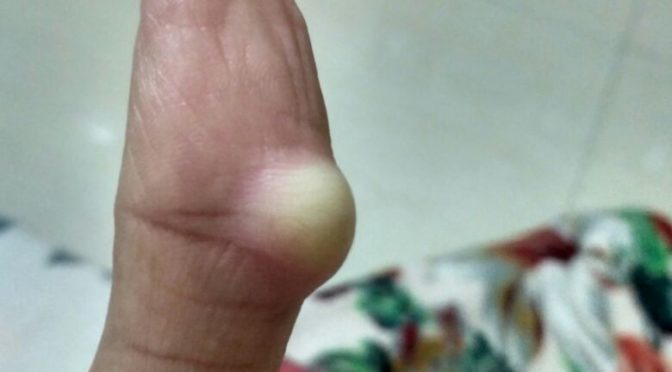
ONE OF OUR CASES OF GANGLION CYST CURED WITH HOMEOPATHIC MEDICINES WITHOUT ANY SURGERY

BEFORE HOMEOPATHIC TREATMENT

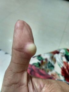
GANGLION CYST ON THUMB
GANGLION CYST ON THUMB, BEFORE TREATMENT

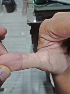
AFTER HOMEOPATHIC TREATMENT

GANGLION CYST OF THUMB RESOLVED AFTER HOMEOPATHIC TREATMENT

TINEA INFECTIONS DERMATOPHYTOSIS

Tinea is the term used to describe a group of contagious skin infections caused by different type of fungi.

Dermatophytes are a group of organisms that are able to breakdown the skin tissues.

Dermatophytosis is mainly caused by microsporum, trichophyton and epidermophyton.

These dermatophytes grow best in warm and humid environments.

DIFFERENT TYPES OF TINEA INFECTIONS

It affects many areas of skin and depending on thier location and fungal infection it has different names.

Common type of Dermatophytosis depending on the sites are as follows

TINEA CAPITIS

Scalp ringworm causes itchy patches on the head, it can leave bald spots, usually affects children.

TINEA BARBAE

It is the infection of the skin especially beard area and moustache and is usually seen in men.

TINEA CRURIS

It is also known as Jock’s itch. Infection of groin, vulva, inner thighs , buttocks.

TINEA CORPORIS

When Dermatophystosis presents on the parts of body like trunks, hands, feet etc.

TINEA UNGUIUM

It is infection of one or more finger nails or toe nails.

TINEA PEDIS

It is also known as Athlete’s foot, causes infection of the foot or inter digits of foot.

CAUSES OF TINEA INFECTIONS

Dermatophytosis are the group of organisms that are able to break down the skin tissue. The fungi reside in the soil and are involved in decomposition and can infect the living organisms.

Dermatophytes are transmitted from person to person and some are adapted to animals.

Dermatophytosis is mainly caused by microsporum, trichophyton and epidermophyton. These dermatophytes grow best in warm and humid environments.

It can be easily transmitted through skin to skin contact with an infected person or animal or through indirect contact with an object or surface that an infected person or animal has touched.

Bathroom floors, bathmaths, towels, showers and communal bathing, swimming and changing room areas are common source of infection.

On contact with skin, the dermatophyte spread to the surface layers of the skin.

SYMPTOMS OF TINEA INFECTION

Symptoms of the fungal infections vary depending upon the infection and affected tissues and area of the body.

The skin lesions are usually characterised by redness, inflammation that is more severe at the edges with raised and thick edges, it is usually seen that the central area is clear.

SYMPTOMATIC REPRESENTATION OF TINEA DEPENDING ON THE AFFECTED SITE

TINEA CAPITIS

Tinea Capitis is also known as scalp ringworm. It usually affects children. Seen in children with infection of hair and scalp. The infection is characterised by scaly, irregular or well marked redness with patchy hair loss, the patchy areas are dry with loss of hair and minimal inflammation.

TINEA BARBAE

The dermatophytic infection that involves hair and skin of the face that is the beard and moustache. The lesions may include scaling with pus along with redness.

TINEA FACIEI

It is seen on non beard parts of the face.

TINEA CRURIS

This is an acute to chronic infection of the groin and adjacent areas like inner thighs, butocks. The symptoms usually imclude burning, severe itching and redness with scales, raised edges, sharply demarcated borders with clear centre. Skin becomes dark in the centre as the lesion progreses.

TINEA CORPORIS

Tinea Corporis occurs on the trunk, hands and feet. The infection usually tends to spread to other areas. The lesions are usually pink to red or scaly and annular with slightly raised and sharp margins with clear centre. Sometimes lesions may present with pus.

TINEA UNGUIUM

Tinea Unguium is infection of nails characterised by thickened opaque, a discoloured or deformed nails. The nail may be separated from nail bed. Toe nails are more affected than finger nails.

TINEA PEDIS

Tinea Pedis is seen between the digits of feet. Commonly called as athlete’s foot. There is dryness, fissures and scales, moist lesions in some or all spaces. The lesions may be present with scaling of the soles with inflammation and dryness or redness.

ROLE OF HOMEOPATHY IN TINEA INFECTIONS

Homeopathic treatment is very beneficial and effective mode of treatment that boosts the bodys immune process to fight and eradicate fungal infections.
As the infection gets older, it tends to get more resistant to any form of treatment, once external applications are used for months together the infection becomes resistant. Homeopathy plays an important role in such cases.

INDICATED HOMEOPATHIC REMEDIES

SULPHUR

Sulphur is very beneficial homeopathic medicine for various skin diseases. In cases of ring worm lesions sulphur is quiet useful.
The patient presents with intense itching and burning in erruptions. Indicated in cases of infections that have been suppressed by local medications of various kinds.

PSORINUM

Psorinum is one of the indicated remedy for skin infections. Mainly used for ringworm infections of scalp and bends of joints. There is severe itching in the affected area. Itching worse due to warmth of bed. Indicated in tinea capitis. The hair appears dry, rough, and lustreless. Profuse sweating with offensive body odour throughout the body.

SEPIA

Indicated in case of fungal infections appearing in isolated parts of body. Infections can occur on any part of the body, but common location is bends of knees and elbow. The erruptions are accompanied by itching and scratching, no relief from scratching. High senditivity towards cold air.
Irritable, indifferent towards family and friends.

THUJA

Thuja is indicated in skin infections. Main action is on skin. Indicated in case of dermatophytic infection where the erruptions are on the covered parts of the body, worse from scratching, very sensitive to touch. Worse at night from heat of bed, from cold damp air. Scratching and cold bathing makes the condition worse.

GRAPHITES

Graphitis is one of the indicated remedy for jocks itch Tinea Cruris with recurrent episodes. Indicated where the skin is dry, rough with severe itching. Sometimes moist crusty erruptions are seen on the parts. There is oozing of sticky exudation. Graphitis is effective for fungal infection around vulva.

ARSENIC ALBUM

Arsenic album is beneficial in Tinea Capitis. The scalp shows bald spots with intolerable itching with intense burning. The symptoms are usually worse at night. Arsenic alb helps to reduce itching and burning and also helps in regrowth of hair on bald spots.

SILICEA

Silicea is indicated in Fungal infections and dermatophytosis in Delicate pale waxy patient. Scars are painful. Eruptions itch only in the day time and evening. Crippled nails. Indicated in case of Tinea Pedis with offensive sweat, deep cracks between toes with intense itching and burning. Painful foul smelling ulcers of the foot.

PERTROLEUM

Petroleum is indicated in case of Tinea Pedis. Helps to drive away infection of foot. Presents with offensive sweat. Deep cracks between toes with intense itching and burning of skin. Crippled and brittle nails.

TELLURIUM

Tellurium is indicated in Dermatophytosis with lesions on large parts of the body. Usually it covers a broader area of skin. Presents with excessive itching and stinging sensation. Itching day and night. Cold air worsens the itching. Offensive odour from the affected area.

CHALAZION

WHAT IS CHALAZION?

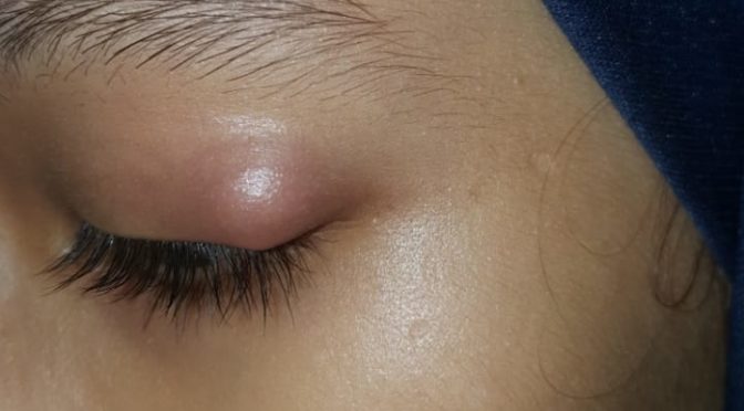
Chalazion is a small, usually painless lump or swelling that appears on eyelids as a result of cyst formation due to blockage of duct of Meibomian gland or rarely of Glands of Zeis. It can appear on upper and lower eyelids. It can be single or multiple and can affect both the eyelids of both the eyes at same time.

CAUSES AND RISK FACTOR

CHALAZION is caused by a blockage in duct of one of the tiny Meibomian Gland of upper and lower eyelid or due to infection or inflammation of Meibomian Gland and rarely of Gland of Zeis in which it is usually secondary to Stye.

Meibomian Glands are responsible for secretion on Meibum which is oily in nature that traps tear within eye by forming thin layer above surface of secreted tear, thus retaining it in eye by not letting it spill out and also reduces evaporation of tears thus playing a major role in keeping eye moist. There are around 50 mebomian glands in upper eyelids and 25 mebomian glands in kower eyelids they are situated on the rim of eyelids on Tarsal plate

Glands of Zeis are sebaceous glands that open up in hair follicle of eyelash they secrete sebum to keep eyelids and eyelash soft and supple, it also plays part in providing lubrication. In this gland the chalazion formed is usually secondary to stye and is not too frequent.

It is more common in people with following risk factors

  • Chronic Blepharitis
  • Seborrhea
  • Rosacea
  • Long term inflammation of eyelids.
  • Infective conjunctivitis.
  • Person wearing contact lenses.
  • Those who have frequent exposure to pollution, smoke, dust etc.
  • Those using Kajal or Surma.

SYMPTOM OF CHALAZION

  • Painless lump or swelling in upper and lower eyelid usually visible or palpable.
  • It can affect both eyes at the same time.
  • Usually they are well defined round with sharp margin and whole surface of sphere is easily palpable if held netween fingers with eyelids gliding over it.
  • There can be one or multiple chalazia.
  • Sometimes in cases of multiple the chalazia, due to frequent secondary infections, they may mate and overlap adjecent chalazia.
  • In mated and overlaping multiple chalazia it becomes necessary to rule out malignancy.
  • Symptoms depend on its size larger chalazia may cause disturbance in vision due to obstruction of viewing fleid.
  • In some cases it may cause nystagmus.
  • If there is secondary infection then it  might be red and inflammed in appearance and may also occasionally be painful.
  • May complicate and show blepheritis as well.

DIAGNOSIS

Sometimes Chalazion may be confused with external or internal Stye.

DIFFERENCE BETWEEN STYE AND CHALAZION.

EXTERNAL STYE

Is caused due to infection in the area of eyelash follicle or sweat gland.

INTERNAL STYE

It is caused due to infection of a meibomian gland.

Difference between chalazia and stye is that stye are painful but chalazia usually aren’t and chalazia may develop after stye.

By physical examination or by asking the patient if there is pain so that it can be differentiated whether it is stye or chalazion. Although chalazion with secondary infection may be painful, so proper evaluation is required in all doubted cases, by checking the location and palaption helps to diagnose properly.

Chalazia those are abnormally large or has abnormal texture of skin over it or is fixed and skin over it is not freely gliding or they are recurring or multiple or overlaping or suspicious with other unusual characteristics should be investigated for malignancy.

Differential diagnosis includes

  • Stye
  • Mebomian adenoma
  • Carcinoma of sebaceous gland
  • Sarcoid Granuloma
  • Foreign Body Granuloma.

TREATMENT

Chalazion may disappear by its own just use a warm compression to your eyelid it can reduce the swelling. If it does not reduce then necesary treatment is essential.

ROLE OF HOMEOPATHY:

Under conventional mode of treatment, surgical removal of chalazion is the only option. which has its own comolications like dryness of eyes and irritation of eyes due to scar formation at site of excision and also it damages and makes mebomein glands non functional forever.

However, In homeopathy chalazion is treated in a non invasive manner with gentle and effective approach.

Few indicated homeopathic remedies in Chalazion are

APIS MELLFICA

Presents with swollen eyelids, redness, intolerance of heat and touch especially right sided with burning and stinging sensation.

PULSATILLA NIGRICANS

Patient presents with itching and thick profuse discharge, Worse in warm room better in open air. There is dimness of vision swelling of eyelid and patient wishes to rub or wipe eye constantly.

STAPHYSAGRIA

Indicated in Recurrent chalazion, better by warmth application. Sunken eyes , heat in eyeball, feel dry despite lachrymation.

THUJA OCCIDENTALIS

Indicated in case of Chalazion. Eyelids stick together, usually left sided, eye lid feel heavy as lead and may be dry and scaly. Bluring of vision.

GRAPHITES

Indicated in case of chalazion, recurrent stye, conjunctivitis, red and swollen eyelid with much dryness it is good for blepharitis too. Indicated remedy for eruption on lids that become hard over a period of time.

CALCAREA FLOURICA

Indicated in case of chalazion. Gland enlarged and become stony hard useful in patient has had chalazion for a very long time. Swelling of eyeild with pain.

CONIUM MACULATUM

Indicated in case of chalazion. Frequently used remedy for stye and chalazion which have a tendency to become stony hard over a period of time for chronic and long standing complaints.

HEPAR SULPHURIS

I have found this remedy effective in almost allthe cases of chalazion when given in 30 potency with frequent repetition 2-3 times a day for a prolonged period of time and then chalazion starts showing signs of inflamation after which the medicines should be stopped and soon we see the chalazion gradually is resoved in few weeks after the medicines were stopped.