All posts by Dr Deepan

Dr DEEPAN P SHAH MD(HOMOEOPATHY) SPECIALISED HOMEOPATHIC PHYSICIAN AND SURGEON

CARPEL TUNNEL SYNDROME CTS

CARPEL TUNNEL SYNDROME CTS is a condition where in the compression symptoms arise due to bundle compression of structures that pass within carpel tunnel affecting the the Median Nerve.

WHAT IS CARPEL TUNNEL?

CARPEL TUNNEL is normal anatomical structure in our body found in every normal individual. It is formed by carpel bones forming a groove as its floor on dorsal side of hand and flexor retinaculum forming the flat roof of carpel tunnel on palmar side of hand.

It provides attachment and passage at the wrist level for the structures to pass through arm to hand. Muscles related to flexing movements of finger pass through this tunnel alomg with median nerve

Structures Passing Through Carpel Tunnel

  • Flexor Digitalis Superficialis – 4 tendons
  • Flexor Digitalis Profundus – 4 tendons
  • Flexor Pollicis Longus – 1 tendon
  • Median Nerve
  • Flexor Carpi Radialis not exactly pass within the carpel tunnel but it traverse through Flexor Retinaculum that is forming the roof of carpel tunnel.

CAUSES OF CARPEL TUNNEL SYNDROME

Anything that exerts pressure to median nerve giving rise to nerve compression symptoms cause carpel tunnel syndrome. In most cases the cause remains obscure and are idiopathic. Any inflamatory process or water retention or metabolites deposition within tissue may cause increase in volume of structures and total content within the carpel tunnel and will cause in pressure of whole bundle which may cause Carpel tunnel due to increased bundle pressure, similarly the adjecent structures to carpel tunnel if inflamed or injured or any overgrowth of it, may exert pressure on carpel tunnel and median nerve within giving rise to carpel tunnel syndrome. Mechanical reasons like wear-tear and injuries are mostly regarded as major factors with ageing and certain genetic, structural and physiological anomalies are known to increase the risk of Carpel Tunnel Syndrome.

Age and Gender

  • Age – Ageing plays a major role in development of CTS its more commonly found in age above 40yrs
  • Gender –  Female sex is more prone to this condition compared to male.

Mechanical

  • OCCUPATIONAL – Work related frequent repetitive forceful pressures jerk and vibrations on hand on regular basis where in there is no sufficient time given to repair the wear and tear and the damage tend to accumulate over time.
  • POSTURAL HABBITS such that it exerts unusual strain on hand and wrist on regular basis.
  • INJURIES to wrist distal part of forearm and wrist. Fractures involving radius ulna carpel and metacarpel bones like Colle’s Fracture, Boxer’s Fracture etc.

Genetic and Systemic 

  • Prediabetics and Diabetics
  • Obesity
  • Metabolic Syndrome
  • Hypothyroidism
  • Arcomegaly
  • Amyloidosis
  • Rheumatoid Arthritis
  • Gout
  • Tumours
  • Hereditary Neuropathy with Susceptibility to Pressure Palsy – Carcot Marie Tooth Syndrome casused due to mutation in gene HS3TC2
  • Narrow Carpel Tunnel
  • Certain drugs like Corticosteriods or Oestrogen therapy.

SIGNS AND SYMPTOMS OF CARPEL TUNNEL SYNDROME

  • Paraesthesia in Index finger, Middle finger, Thumb and the lateral half of  Ring finger
  • Myalgias or neuralgia in forearm upwards to arm, less commonly in wrist and hand
  • Loss of strength of grip and Loss of Manual Dexterity
  • Atrophy of muscles at the base of thumb visible on proximal, thumb-related quadrant of palm (ie proximal lateral quadrant/upper outer quadrant).

HOMEOPATHIC MEDICINES FOR CARPEL TUNNEL SYNDROME CTS

  • HYPERICUM
  • CIMICIFUGA/ACTEA RACEMOSA
  • AGARICUS MUSCARIOUS
  • ACTEA SPICATA
  • PLANTAGO
  • BELLIS PERENIS
  • ARNICA MONTANA
  • GELSEMIUM SEMPERVIRENS
  • TARRENTULA HISPANICA
  • GNAPHALLIUM

SHIV-LING – VEDIC NUCLEAR REACTOR MODEL?

SHIV LING and NEUCLEAR REACTOR

Shiv Ling Vedic Model of Nuclear Reactor?

Shivling is found on earth on almost all continents with its reference found in vedic literature and archeological findings date it back to thousands of years, back in vedic era.

I was always confused when I got to know that many people obscenely depicting Shivling falsely as some body part of Shiv due to word “Ling”, where as its a formless god for vedic culture and symbol of creation, maintenance and destruction.

Its well said by Mahatma Gandhi that “Each one prays to god in his own light” – So this also explains that it reflects one’s knowledge, character and personality that how he percieves everything.

Vedas are compilation of poetic verses with vast knowledge of science, society and universe, its a bare act of nature. Each verse is being crafted with extreme efforts to preserve and carry forward to next generations, numerous messages in shortest, most perfect and meaningful sentence. Its compiled in poetic verses as there are many dimensions to each aspect of nature that simple language cannot explain and its not something only to be undertood but its has more dimensions to it, as it needs to be felt and realised to understand it and so the poetic verses, symbolisation and personification are the best ways that can carry the message in depth to next generation.

Its difficult for us to understand and  explain lord shiva like the way Vedas do and the understanding Vedas give to one who meditates each shlok in depth of it. But still if one has to attempt to explain, then the frequently used explanation is that Shiva is Formless, Omnipotent, Omnipresent, All pervaded by him, Creator, Maintainer, Destroyer, Nothingness, Final Emancipation, The Auspiciois One and many more attributes,  to understand each of the above aspects better, we need to understand aspects of nature, physics and emotions with all its dimensions first.

Shiva is mostly worshiped in form of Shivaling and only personified form that it is worshiped is Nataraj form where its also called the dance of Lord Shiva, now this personified representational form as per worlds largest particle physics laboratory (CERN) is beautiful dipiction of harmonious motion of electron with respect to other sub-atomic particles and so they have placed large Natraj Idol at the entrance of CERN’s Large Hadron Collidor and many physics and religious scholar after going through Vedic literature and current science findings believe Shiva to be depiction of some sub-atomic field that makes the basic fabric of whole universe, so recently found Higgs Bosson was termed as God-Particle.

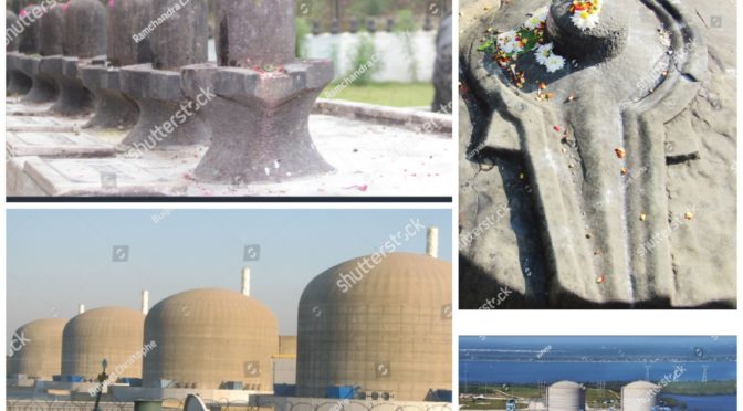
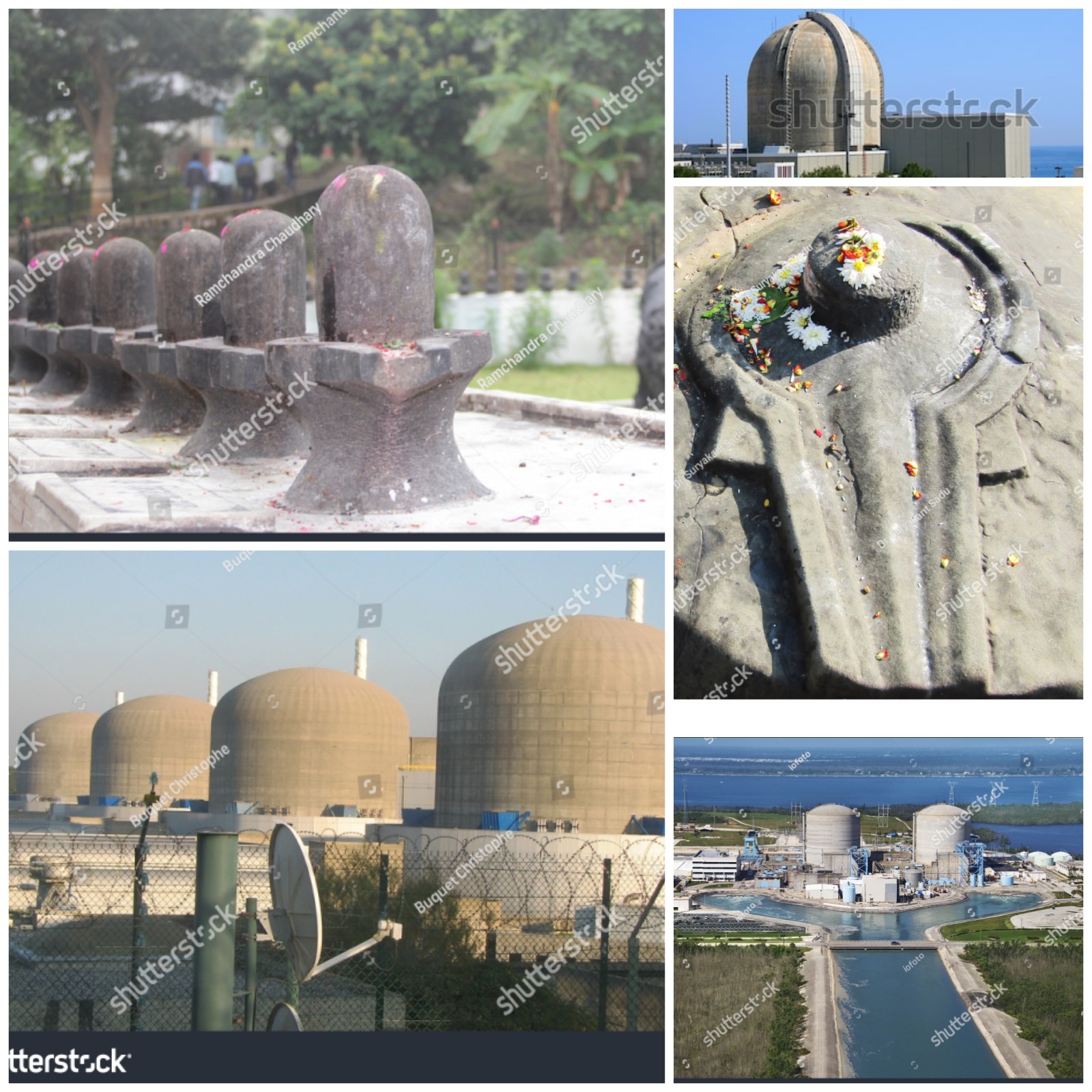
Now if we further relate one of the representational form of Shiva, that is the  Shiv Ling, then its dome shaped with water continiously being dropped over it with channels around it to drain the water off, Shiv ling are almost always found near large water resource like river or sea, so that it get continous water supply and draining. The drained water is prohibited to touch or consumed as its regarded unsafe for health.

Now of we compare Shivling with Neuclear Reactors, Nuclear Reactors are dome shaped, needs continious robust water supply to keep it cool and are built near large water resources like sea, with channels of water drainage. Also certain wastes from Neuclear plant are radioactive and dangerous to come in contact with.

And also looking at above explained aspects and belief of scientists about the Shiva, “The Shiv-Ling as Neuclear Reactor Assumption” has got good chance to not to get rejected as probable model of Neuclear Reactor or even something related beyond it.

DID I DECODE SWASTIK? CHEMICAL FROMULA THAT GIVES LIFE AND GIVES MOTION TO BREATH?

SWASTIK CHEMICAL FORMULA FOR BREATH AND LIFE
SWASTIK – CHEMICAL FORMULA OF LIFE H4CO4 in SANSKRIT LANGUAGE “SWAAS” = BREATH “TIK” = MOTION or LETTING OUT

SWASTIK is regarded as very auspicious symbol in Hindu religion with its origin in Vedic culture

Vedas are the oldest known literary work on planet Earth and are considered as authorless. In Hinduism Vedas are regarded with highest order, amongst all religious and spiritual scriptures. All other scriptures are derivative of vedas or correspond to ideas of vedas, it is considered to be the bare act of nature.

Swastik symbol is found in almost all continent in excavation work and its considered to be the oldest symbol ever found on earth with its references in almost all the cultures around the world. Especially in Hinduism Swastik is consodered very auspiciois and it is used in each and every religious and spiritual activities.

The best way to pass on data, is in easy symbol forms, so that civilization, if not knowing a particular language, even then they can decode and understand.

Because of all these factors Swastik always made me wonder what it must be encoding within, as vedic culture is known to be rich in knowledge and its known to forward knowledge in form of poetic verses and symbols.

So, while studying carbon and carbon compounds, I came across Atoms of 3 different elements

  • Carbon atom which has valency of 4
  • Oxygen has valency of 2 and
  • Hydrogen with valency of 1

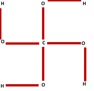
These atoms of these 3 elements if placed in Swastik with Carbon in center of Swastika, Oxygen at the turn of each limb of Swastika and Hydrogen at the end of each limb of Swastika forms a complete chemical code of Swastik H4CO4

Now wether they actually meant this or something else? Is it symbol for chemical formula that they made or they wanted to represent something else?

So, now if we try to further decode Swastik we can break word “SWAS-TIK” into two simple words

  • “SWAS”  means breath and
  • “TIK” means motion or letting out in sanskrit language.

LINK OF SWASTIK CHEMICAL COMPOUND IN RESPIRATORY PROCESS

Now further we are able to match this chemical code with the meaning of these two words “SWAS” and “TIK”

Swatik is highly unstable compound on planet earth as its atmosphere and  its atmospheric pressure doesnt let it thrive, as it requires extremely high pressure to thrive, otherwise it decomposes soon into the nearest stable compound.

4HCO4 decomposes into Carbonic Acid and Water as shown in chemical formula below.

4HCO4 ➡  CH2O3 + H2O

CH2O3 has important role in respiratory process. It is the only acid secreted by lungs in the form of gas and it is described as “Volatile Acid” or “Respiratory Acid”.

So is swastik related to a very unstable intermediate osscilatory substance which though not actually comes into form but remains as intermediate exchange form and plays a major role in respiratory process and constant respiratory osscilation?

??? SO DOES SWASTIKA MEANS THE COMPOUND THAT GIVES MOTION AND OSSCILATION TO RESPIRATION!!!

TRIGGER FINGER

TRIGGER FINGER or TRIGGER THUMB is a condition where in joints of one or more of the digits(finger/thumb) gets stucked up/locked up at certain postion which is difficult to move and moving it with force may cause popping or clicking sound with pain.

Though it can present in any sex and at any age, it is more commonly found in females around age of 50-60 yrs.

CAUSES OF TRIGGER FINGER/THUMB

Its is also termed as digital Stenosing Tenosynovitis, although there is no predominant inflamation to Tendosynovium but inflamation is found in Tendon Sheath. And also its not comfirmed that inflamatory process has any primary role in its development

Though exact cause behind trigger finger/thumb is not known but the risk factors that tend to increase the incidence of this condition are identified

  • Over straining and over use of hand and fingers espescially activities involving prolonged forceful flexion of digits(fingers/thumb) may be occupational or habbitual routine activities.
  • Frequent injuries – occupational, accidental or even injuries of planned surgery of hand especially ofter surgery for carpel tunnel syndrome
  • Systemic connective tissue disorders
  • Autoimmune diseases especially Rheumatoid Arthritis
  • Hypothyroidism
  • Renal Disease
  • DeQuervain’s Diseases
  • Amyloidosis
  • Diabetes Mellitus and Other Metabolic disorders.

SYMPTOMS OF TRIGGER FINGER/THUMB

  • Though it may involve any digit(thumb/finger) Index finger and Thumb are more frequently involved. One or more digits may be involved.
  • Patient presents with stucked up digit at certain position. It may be at any level from flexed  to extended position usually found at semiflexed position.
  • This locking up may be persistent for a prolonged period of time or may be momentary and recurrent.
  • Aggravations are more commonly experienced at night, especially while holding heavy article with hand or while gripping or applying pressure with fingers or hand.
  • On moving and or forcefully unlocking the stucked finger is bit painful and causes clicking and popping sound
  • In severe cases the finger may be persistently locked for prolonged period of time with constant pain which may also extent to whole hand and wrist.

DIAGNOSIS OF TRIGGER FINGER/THUMB

Diagnosis of trigger finger/thumb is based on clinical symptomatology of the patient where in inflamation or involvement of tendon sheath of flexors is confirmed and excluding probability of other condition like

  • Tumour
  • Sprain of digit
  • Osteoarthritis of metacarpo-phalangeal joint
  • Fracture
  • Tendon Entrapment post-trauma
  • Extensor Apparatus Injury
  • Game Keeper’s Thumb
  • Sesamoid anomaly
  • Dupuytren’s Contracture
  • Focal Dystonia

An injection of lidnocaine in tendon sheath of flexors reduced the pain then it confirms diagnosis Trigger Finger/thumb.

HOMEOPATHIC TREATMENT FOR TRIGGER THUMB

Treatment of trigger finger/thumb is dependent on combination of modalities of physiotherapy, splinting and medication.

HOMEOPATHIC MEDICINES FOR TRIGGER THUMB/FINGER

  • CALCAREA FLOURICA
  • SILICEA
  • THIOSINAMINUM
  • RHUS TOXICODENDRON
  • ACTEA SPICATA
  • THUJA OCCIDENTALIS
  • AGARICUS MUSCARIOUS
  • ARNICA MONTANA
  • BRYONIA ALBA
  • CIMICIFUGA RACEMOSA

REACTIVE ARTHRITIS

Reactive Arthritis was also called Reiter’s Arthritis is RF-negative and HLA-B27 Linked Imflamatory oligoarthritis typical with Enthesitis, accompanied with Inflamatory occular and/or inflamatory genitourinary and other systemic manifestation usually post gastrointestinal or genitourinary infection.

During world war one and two many cases emerged with the Triad of Symptoms viz. Inflamation of Joints, Inflamation of eyes and Inflamation of Uretha. Which drew attention of medical community due to common presentation in many giving it some syndrome like picture. On further investigations it was found out that most of them were exposed to urogenital or Gastro-intestinal infection 1-4 weeks prior to onset of this Triad of Symptoms. This was initially termed as “Fessenger-Leroy-Reiter’s Syndrome” or simply  “Reiter’s Syndrome”. But as the physician Hans Conard Julius Reiter  was involved in attrocities and war crimes with Hitler, so his name was removed and later renamed and termed as “Reactive Arthritis”.

EPIDEMIOLOGY OF REACTIVE ARTHRITIS

  • AGE – It more frequently affects age group of 20-40 years.
  • SEX – It is more common in Males then in Females.
  • ETHNICITY – Due to its association with HLA-B27 it is frequently found in white race compared to dark race as comparatively HLA-B27 occurs more commonly in white population.
  • RISK FACTOR – Person with HIV positive status are more prone to develop reactive arthritis.

SIGNS AND SYMPTOMS OF REACTIVE ARTHRITIS

The onset of symptoms of Reactive Arthritis typically starts 4-35 days after an initial infection of gastro-intestinal system or genito-urinary system.

TRIAD OF REACTIVE ARTHRITIS

Reactive arthritis in most of the cases presents where patient cant – SEE, PEE, climb the TREE! due to following Classical Triad of Symptoms of reactive arthritis

i) OLIGOARTHRITIS

Oligoarthritis involving less than five joints. It may frequently involve knee and sacroilliac joint as well. May present itself in additive pattern where it starts with one joint and add another joints subsequently or it may be migratory in pattern where the set of inflamed joints keep changing by addition and simultaneous substraction of joints involved.

ii) NON-GONOCOCCAL GENITOURINARY INFLAMATION

Inflamation of genitourinary system classically presents itself at the onset of the disease. Not always but in many its typically after initial sexual exposure. It presents as frequent burning micturation, uritheritis, prostatitis, balanitis in men and salpingitis, vulvitis and vaginitis in women.

iii) OCCULAR INFLAMATION

Occular Inflamation may present itself as mild conjunctivitis or uveitis in 75% of cases with gastrointestinal origin and 50% of cases with urogenital  involvement. patients have intermittent irritation in eyes with blurred vision typically commences at onset of disease.

OTHER SYMPTOMS

  • Few patients also presents with peculiar symptom which is specific to reactive arthritis, its Keratoderma Blenorrhagica which are small hard nodule commonly appear on soles occasionally on palms and rarely on other parts of body subcutaneous nodules are not incluced. Even in absence of above mentioned triad of symptom the presence of Keratoderma Blenorrhagica is diagnostic for reactive arthritis.
  • In reactive arthritis; typical to HLA B27 related immunological reactions; involves Entheses that is where skeletal muscles attaches with bones through tendons, where it causes Enthesitis and tendon inflamation especially the tendo-achilles and also fascia in particular Plantar Tendinitis.
  • Occasionally patients also suffer from dactilitis giving finger sausage-like apperance “sausage finger” due to inflamation.
  • Mucocutaneous involvement presents as ulcerative or non ulcerative stomatitis, apthous ulcers and geographic tongue are also seen as presentation of this disease
  • Cardiac involvement causing pericarditis and aortic regurgitation in cases which do no recover soon or if its recurring or progressive.
  • Gastrointestinal manifestation like pain and cramps with frequent semiformed stools with mucous and insome cases blood due to  inflamation and ulcceration in gastrointestinal tract.

Most of the cases of Reactive Arthritis recover within six months, in many cases it keeps comming back time and again and in few it becomes chronic and progressive which may increase risk of severe complications.

COMPLICATIONS OF REACTIVE ARTHRITIS

In chronic progressive and recurring cases the patient may develop following complications

  • Ankylosing Spondylosis
  • Disabling Arthritis
  • Aortitis
  • Aortic Regugitation
  • Conduction defects of Heart
  • Pericarditis
  • Amyloid deposits
  • Immunoglobulin A Nephropathy

CAUSE OF REACTIVE ARTHRITIS

Reactive Arthritis is is HLA B27 linked inflamatory arthritis and enthesitis preceeded by a spell of infection either of genito-urinary system  or gastro-intestinal system by following commonly involved organisms

GENITO-URINARY INFECTIONS ASSOCIATED WITH REACTIVE ARTHRITIS

  • Chlamydia Trachomatis
  • Ureaplasma Urealyticum

GASTRO-INTESTINAL INFECTIONS ASSOCIATED WITH REACTIVE ARTHRITIS

  • Salmonella Spp.
  • Shigella Spp.
  • Campylobacter Spp.
  • Yersinia Spp.

4-35 days after the spell of urethritis or food poisoning by above mentioned organisms the symptoms of reactive arthritis sets in, where the synovial fluid has negative culture ans is free from infection and but the HLA B27 linked  inflamation is thought to be triggered due to

  • Autoimmune reaction due to cross reactivity of micro-organism antigen with joint tissue  or
  • Micro-organism antigenic components that may have settled in joint tissue.

DIAGNOSIS OF REACTIVE ARTHRITIS

Clinically the Reactive Arthritis can be diagnosed with help of Sensitivity and Specificity Guidlines laid down by American College of Rheumatology, for clinical diagnosis with given set of presenting symptom, its as follows

  1. Arthritis > 1 month with Urethritis and/or cervicitis has  sensitivity of 84.3% and specificity of 98.2%.
  2. Arthritis > 1 month with Urethritis or Cervicitis or bilateral Conjunctivitis has Sensitivity of 85.5% Specificity of 96.4%.
  3. Arthritis, Urethritis and Conjunctivitis has Sensitivity of 50.8% and sensitivity of 98.8%.
  4. Arthritis > 1 month, Conjunctivitis and Urethritis has Sensitivity 48.2% and Specificity of 98.2%.

Patients falling in above criteria or those showing just Keratoderma Blenorrhagica without any other symptoms and other suspected cases can be sent for following test for further evaluation.

  • HLA B27 testing
  • Urine routine and culture
  • STOOL Routine and culture
  • Throat swab culture
  • Cervix and Urethral swab culture
  • Erythrocytes Sedimentation Rate
  • C-Reactive Protein Test

HOMEOPATHIC TREATMENT FOR REACTIVE ARTHRITIS

Being an immune mediated systemic reaction that too the one that is triggered with different causative agents and even to same agents different individuals will respond differently.

Though they may have same set of general symptoms like the classical triad of reactive arthritis but intensity of each of the symptom of triad will differ in each individual,

Now this is where the homeopathic individualisation process starts. In Homeopathy we believe that though majority of human genome is the same but the minor variations in gene and the epigenome make the whole lot of difference in various characteristerics of each individual, similarly their immune reaction also varies, so every person should have individualised medicine.

Homeopathic Treatment is based on symptom similarity and individualisation of case based on peculiar symptoms based on which the case is individualised and medicine is selected.

Alternatively as per Homeopathic principle of Genus Epidemicus or pathology based symptomatology there can be disease specific homeopathic medicine derived from common symptomatic representation of a disease condition in a group of population.

Now this can not be the most similimum homeopathic prescription but roughly it can hit the disease condition within an indivudual though not accurate but will yeild some results in most of the cases.

To yield best homeopathic results there can be no generalised common approach for all cases.

But still if we have to attempt common standardised pathology based approach then to give some guidelines on homeopathic approach towards cases of reactive arthritis I have attempted following rough guidelines which may help to give some vision in approach towards such cases.

Its seen that in few case it begins after gastro-intestinal infection and in some case post genito-urinary infection. So this will further guide determining “morbid cause” behind the disease directing us in homoeopathic similimum medicine selection.

Now reactive Arthritis shows a triad of symptom in most of the cases. So this triad helps us to reach to group of medicines with such combination of symptoms.

Intensity, occurance of symptoms and its sequence in triad differs in each individuals. For example

  • In some person urogenital symptoms may be more severe compared to occular symptoms or arthritis symptoms, where as in others arthritis and ocular symptoms would be more severe than urogenital symptoms.
  • Some may not have occurence of  conjunctivitis
  • In some all three triad occur at a time where as in some patients it may occur gradually one after another in different sequence.

All this helps us find out the “seat of disease” in an individual and its degree of affinity towards various organs which can be related to homeopathic medicines during selection process.

Further arthritis may show different pattern like

  • progressive
  • migratory
  • additive
  • symetry
  • predominantly involved joint
  • sequence of joint involvement
  • number of joints involved
  • severity
  • intensity
  • type of sensation and other symptoms

Also similarly symptoms of occular involvement and urogenital involvement should be take in to account in absolute detail. This further helps refine and classify the patient and the respective medicines to be repertorised.

Which other systems and organs are involved like mucous membranes, skin, heart, kidney etc and what type of pathology they are showing like tissue destruction or just inflamation and functional disturbance or tissue lysis with regenerated and degenerative changes this will help to decide what “type of miasm” is underlying wether its psoric, syphillitic or psychotic type pathology.

Certain symptom are very “peculiar” for the  disease and occurs in few individuals like Keratoderma Blenorrhagica eruption, now location of this eruption will further help individualise the case.

Enthesitis – Inflamation of tendo-achilles and plantar fascitis is  “very specific” to the disease but does not occur in all individuals, so if plantar fascitis or inflamation of tendo-achilles if occurs in someine with this disease then it helps further in individualisation of during homeopathic medicine selection.

Other than this the general health and family background should be noted to derive  constitutional types and association of HLA B27 in 75% of this individual further helps in individualisation and homeopathic medicine selection.

COMMONLY USED HOMEOPATHIC MEDICINES FOR REACTIVE ARTHRITIS

  • PHOSPHORUS
  • ARSENICUM ALBUM
  • RHUS TOXICODENDRON
  • BRYONIA ALBA
  • LEDUM PALUSTURE
  • THUJA OCCIDENTALIS
  • ANTIMONIUM CRUDUM
  • ARGENTUM NITRICUM
  • BORAX
  • MERCURIOUS SOLUBILIS
  • PULSATILLA NIGRICANS

FROZEN SHOULDER HOMEOPATHIC TREATMENT

Frozen Shoulder also called Adhesive Capsulitis of shoulder joint is painful stiffness limiting range of motion of shoulder joint caused due to inflammation related pathophysiology in capsule of shoulder joint.

CAUSES AND RISK FACTORS OF FROZEN SHOULDER

CAUSE OF FROZEN SHOULDER

Exact cause of Frozen Shoulder remains unclear but there are many factors that increases the risk and lead to frozen shoulder

RISK FACTORS FOR FROZEN SHOULDER

GENDER

Frozen shoulder is more common in females than in males

AGE

Person above 40yrs of age are more prone to this condition

INJURIES

  • Injury to shoulder joint involving any of its companent especially gleno-humeral joint capsule or rotator cuff.
  • Sports persons typically develop calcifications of tendons around shoulder joint and also in joint structures due to repeated injury and result of prolonged inflamation such persons are at high risk of developing frozen shoulder.

REDUCED MOBILITY

  • Injuries to arm and neck may indirectly affect shoulder joint, due lack of motion in general of that region as a whole and changes pattern of gait and movements of that region.
  • Immobility or reduced mobility post surgerr with general debility and delayed recovery.
  • Immobility post stroke.

MUSCULO-SKELETAL DEFECTS

  • Certain congenital structural anomaly of Musculoskeletal framework which has influence on movement, gait and weight distribution pattern on shoulder joint in abnormal pattern, which may put the person at risk of develooing Frozen Shoulder.
  • Kyphosis or Scoliosis or other congenital or acquired spinal anomaly especially those above thoracic spinal level may increase risk of frozen shoulder.
  • Patients with cervical spondylosis are at risk of developing frozen shoulder.

RHUEMATIC DISORDERS

OTHER SYSTEMIC DISEASES

SYMPTOMS OF FROZEN SHOULDER

  • Pain in affected shoulder aggravates on movement
  • Loss of rang in motion of affected shoulder joint

Frozen Shoulder is divided into 3 stages

1) Freezing Stage

In Freezing Stage there is pain in joint which aggravates on motion and he range of motion gradually starts decreasing.

2) Frozen Stage

In Frozen Stage the pain is not much as compared to freezing stage but the range of motion is completely diminished so much so that patient can barely move the joint or cant move at all.

3) Thawing Stage

In Thawing Stage pain subsides and range of motion gradually starts improving and complete recovery is established in few weeks.

If it is acted upon quickly in freezing stage with intervention of physiotherapy and proper medication the progress of the disease is arrested and soon resolves completely.

DIAGNOSIS OF FROZEN SHOULDER

On clinical presentation of symptoms of the patient frozen shoulder can be diagnosed and radiological tests like X-rays, CT scan and MRI may be suggested inrequired cases to find out extent, severity of pathological condition within and also to rule out any other pathological condition within or around shoulder joint.

If along with shoulder joint if there are other joints involved simultaneously or frequently different joints in past then it becomes necessary to rule out other underlying systemic conditions like Rheumatoid ArthritisGout, Ankylosing Spondylosis, Systemic Lupus Erythematosus etc and for that your doctor may suggest blood tests like RA factor, ESR, CRP, ANA, S.uric acid and HLA B27 (where there is severe spinal involvement along with shoulder). Vitamin D should also be checked wether its in optimal range for better recovery.

TREATMENT OF FROZEN SHOULDER

Treatment of frozen shoulder is based on combination of both Physiotherapy and Medicines.

Physiotherapy plays a major role in cases of frozen shoulder. Physiotherapist takes support of certain modalities, recomends certain exercises and manual physical intervention where in he may exert mild force or pressure along and within the axis of movement of shoulder joint and try to open up the freezed shoulder and gradually retain its laxity and mobility.

Pain may temporarily increase during and  after physical intenvention but with regular recomended exercise and medicines it finally subsides and joint regains its complete range of axis of movement subsequently.

HOMEOPATHIC MEDICINES FOR FROZEN SHOULDER

If the frozen shoulder is without any underlying condition specific medicines will work well but if it is secondary to some underlying condition like autoimmune diseases or metabolic disorders or Infections or some other systemic or acute or chronic complaints then constitutional trearment along with initial acute specific remedy may be required.

List of Common Homeopathic Remedies Used in Frozen shoulder

  • Calcarea Flourica
  • Silicea
  • Calcarea Phosphorica
  • Thiosinaminum
  • Rhus Toxicodendron
  • Bryonia Alba
  • Arnica Montana
  • Plantago Major
  • Belladonna
  • Ledum Palusture

LUMBAR SPONDYLOSIS

Degenerative changes in vertebral bodies and intervertebral disc at the level of L1 to S1 in vertebral cloumn affecting lumbar region of spine is called Lumbar Spondylosis.

It is characterised by degenerative changes of Lumbar vertebral bodies like osteophyte lippings and spur formation, degeneration of  intervertebral disc, narrowing of intervertebral space, Intervertebral disc bulging, intervertebral disc herniation.

It can also be called Osteoarthritis of Lumbar Spine.

CAUSES OF LUMBAR SPONDYLOSIS

Lumbar Spondylosis may be caused due to many factors that can be broadly classified into Mechanical, Congenital or Acquired Anatomic anomalies, Metabolic, Immunologic, Genetic, Nutritional, Ageing and Septic reasons.

Most common of all are ageing and mechanical reasons like excessive wear and tear due to wrong postural habbits, type of work like driving for long duration regularly, sitting for long hours, lifting weights or working with bent posture etc. that exerts much pressure on lumbar spine on regular basis so as to not giving much time to recover again and resulting into accumulation of wear and tear finally damaging the spine and causing lumbar spondylosis.

Metabolic problems like Hyperuricemia/Gout where high level of uric acid in blood damages lumbar spine also this uric acid may get crystalised to form monosodium urate monohydrate and settle in the intervertebral joints and damage them.

Immunologically mediated chronic inflamation in lumbar spine like in Rheumatoid Arthritis, Chickenguniya, Systemic Lupus Erythematosus etc.

Genetic Complaints like Ankylosing Spondylosis etc

Vitamin D deficiency causes reduced calcium assimilation resulting into reduced bone mineral density causing weak bones easy to damage. Insufficient nutrition compared to requirement, which prevents repair work of regular wear and tear of lumbar spine, due to which damages tend to get accumulated resulting into lumbar spondylosis.

Congenital or acquired Anatomical anomalies in musculoskeletal framework that may cause abnormal posture or abnormal weight distribution which results into pressure on spine in wrong fashion gradually facilitates development of Lumbar spondylosis.

Septic Conditions of spine like Tuberculosis of spine may result into lumbar spondylosis due to tissue lysis as result of infection.

SIGNS AND SYMPTOMS OF LUMBAR SPONDYLOSIS

  • Pain and Stiffness  in lower back or Lumbar Region.
  • Sciatica due to compression of nerve roots of sciatic nerve.
  • Pain stinging pain in lumbar spine
  • Pain radiating from lumbar spine to lower limbs
  • Paraesthesia in lower back genitals or lower limbs
  • Cramps calfs, thighs and other muscle and muscle groups of lower limbs
  • Difficulty in walking and maintaining posture and balance.
  • Lack of control on urinary spincter or anal spincter resulting into either retention of urine or bowel or involuntary expulsion of urine and bowel
  • Sexual debility Tabes Dorsalis

COMPLICATIONS OF LUMBAR SPONDYLOSIS

Myelopathy – When there is damage to spinal cord it causes Myelopathy.

Raduclopathy – compression of nerve near nerve root where it emerges out of spinal cord.

Vertebrobasilar insufficiency – Vertebral artery insufficiency due to its occulusion while it passes through vertebral foremen sue to degenerative changes for lumbar spine causes vertebrobasilar insufficiency leading to death of condrocytes of intervertebral disc and osteophytes starts accumulating and depositing.

DIAGNOSIS OF LUMBAR SPONDYLOSIS

  • Clinical symptoms if indicate towards Lumbar spondylosis it can be then confirmed with X ray.
  • MRI and CT scan gives us better imaging which helps  judge and evaluate the extent of damage and condition
  • Myelograph is dye injected while Xray and CTscan this gives detailed shadow to understand even better
  • Electromyography helps evaluate nerve involvement in spondylosis.

HOMEOPATHIC MEDICINES FOR LUMBAR SPONDYLOSIS

  • HYPERICUM PERFOLIATUM
  • CALCAREA CARBONICA
  • GNAPHALLIUM
  • CALCAREA PHOSPHORICA
  • MAGNESIA PHOSPHORICA
  • ARNICA MONTANA
  • NATRUM SULPHURICUM
  • RHUS TOXICODENDRON
  • SILICEA
  • MEDHORRINUM

CERVICAL SPONDYLOSIS

Our spine is classified into cervical, thoracic, lumbar and sacral regions.

Cervical Spondylosis is condition where in vertebra and intervertebral discs of cervical region undergoes degeneration. 

It is characterised by thinning of intervertebral discs, reduced intervertebral space, osteophyte lipping, spurs formations,  herniation of intervertebral disc, nerve compression etc.

It can also be called osteoarthritis of cervical spine.

CAUSES OF CERVICAL SPONDYLOSIS

This degeneration can be due to various mechanical, immunological, infective, metabolic, genetic, nutritional and age related reasons.

It can be due to one or combination of than one ot the above reasons. Most of the cases are due to ageing and mechanical wear and tear related to abnormal physical exertion and postural habbits.

It is commonly seen in people assuming wrong posture for long hours like lying down with head placed on huge pillow or watching mobile phones or reading books with tilted head for long hours, staying on computer workstation with an arm stretched on mouse or key board for long transfers weight on neck.

When such postural habbits are prolonged for few hours to days its starts inflamatory process in cervival spine and if still prolonged for months to years the prolonged inflamation and mechanical wear and tear results into degeneration of spine.

Many genetic and immune mediated conditions like Rheumatoid arthritis, Ankylosing spondylosis, Systemic Lupus Erythematosus, Psoriatic Arthritis etc may result into prolonged chronic inflamation of spine ingeneral and gradual degeneration of cervical spine as well resulting into cervical spondylosis.

Metabolic reasons like hyperuricemia may result into deposition of monosodium urate monohydrate crystals into joint spaces in cervical spine resulting into subsequent erosion and degeneration of spine causing cervical spndylosis.

Certain infections in spine like tuberculosis results into degeneration of spine and spondylosis and when it affects cervical region on spine it causes cervical spondylosis.

With ageing there is depletion of anabolic hormones and other factors required for quick repair process which results into slow repair process compared to daily wear and tear and damages tends to accumulate and gradually resulting into erosion and degeneration of spine.

Nutritional deficiencies arised due to lower intake compared to requirement, resulting into lower calcium vitamin D and many other nutrients which not only slows down the repair process to built up damages but also gives rise to low bone mineral density resulting into erosion and degeneration of vertebral bodies.

SIGNS AND SYMPTOMS OF CERVICAL SPONDYLOSIS

In initial stages it starts with occasional stiffness and pain in neck lasting few minutes to hours after exertion gradually it starts persisting with pain on extreme range of axis of movement of neck then later even on smaller axis or range of motion of neck patient starts feeling stiffness or pain or discomfort in neck

If not taken care the nerves originating from cervical plexus which emerge out from cervical spine they start getting compressed causing myalgia paraesthesia in neck which may radiate to shoulder and extend upto arm and upto tip of fingers.

In severe cases of cervical spondylosis patient may also experience vertigo nausea vomitting complete loss of balance with pain in neck and gastric derangement as concomittant symptoms

COMPLICATIONS OF CERVICAL SPONDYLOSIS

CERVICAL SPONDYLOTIC MYELOPATHY (CSM) – It is caused due injury to spinal cord due to cervical spondylosis.

CERVICAL SPONDYLOTIC RADICULOPATHY – In this the nerve gets pinched and compressed near the nerve root shortly after it leaves spinal cord.

VERTIBROBASILAR INSUFFICIENCY – When vertebral artery which is passing through vertebral formen gets occluded and deprives chondrocytes of intervertebral disc from circulation as a result the die and weaken intervertebral disc and osteophytes starts settling in.

DIAGNOSIS OF CERVICAL SPONDYLOSIS

Cervical Compression Test – When the neck is tilted laterally and applied downward pressure patient feels pain on ipsilateral side in neck or shoulder , its not conclusive but indicative and qualifies case for further radiological investigations

Lhermitte’s Sign – Electrict Shock like pain on flexion of neck

These patients general show reduced range of motion of n

Based on clinical symptoms patient may be sent for X ray  for ascertaining the diagnosis.

MRI and CT Scan helps to further find out extent and severity of damage to spine.

Myelography is done with dye injection in spinal cord during CT or X ray for more detailed radiological imaging

Electromyography and Nerve Conduction Test helps to find out involvement of nerve and nerve damage and extent of nerve injury.

TREATMENT FOR CERVICAL SPONDYLOSIS

Guidlines to patients on maintaining correct posture, avoid jerk and strain to secure neck is of utmost importance in management of cervical spondylosis patients.

Proper calcium intake sufficient exposure to sunlight for vitamin D. Increased protein, vitamin B12 and iron intake.

Exercise like pranayam and walk helps stimulate hormone secretion and thus facilitating absorption and assimilation of nutrients required for repair and rebuilding the worn and damaged tissues and to increase bone mineral density.

Mild gentle exercise of neck helps increase local blood flow and keep tissue supple and stimulate its growth and strenght but if not done under proper guidance of qualified physiotherapist it may further injure the already damaged tissue. So if proper qualified physiotherapist is not available to guide its safer bet not to exercise neck region involving cervical spine on your own and giving it complete rest and let it recover on its own while still continueing with walk and pranayam regularly.

HOMEOPATHIC MEDICINES FOR CERVICAL SPONDYLOSIS

CIMICIFUGA RACEMOSA / ACTEA RACEMOSA

RHUS TOXICODENDRONE

ARNICA MONTANA

PLANTAGO MAJOR

HYPERICUM PERFOLIATUM

TUBERCULINUM

BELLADONNA

CALCAREA PHOSPHORICA

BELLIS PERENIS

SILICEA

MEDHORRINUM

SWINE FLU H1N1

Swine flu is an infection caused by swine flu influenza virus which is common throughout the population worldwide. The disease affects the respiratory tract of pigs and is transmitted to humans.

THE SWINE FLU VIRUS

The influenza virus is a negative sense RNA virus of the family Orthomyxoviridae with three genera Influenza A, B, C.

Swine influenza is known to be caused by Influenza A subtypes. H1N1, H1N2, H3N1 H3N2. In pigs Influenza A virus are divided into subtypes based on 2proteins on surface of virus the haemagglutinin(H) and the neuraminidase (N). There are various types of subtypes of H and N.
Every year new strains of virus evade host defenses.
Another characteristic of type A influenza is that the virus has segmented genome with eight single stranded RNA segments. When the host cell is infected with more than one influenza virus, the genes have the opportunity to get ressorted and produce a different strain altogether. The antigenic shift is responsible for pandemic of influenza which has been observed in the past. Virus acquires the ability to not only infect but spread within the human host.

Influenza B viruses are only known to infect human and seals causing influenza

Influenza C virus infect both human and pigs and does not infect birds.

HISTORY OF SWINE FLU

Swine origin influenza A H1N1 virus was first recognised in the border area of Mexico and United states in April 2009 and during the span of 2 months and was spread with travellers wordlwide resulting in 1st influenza pandemic since 1968. Around mid 20th century identification of influenza sub types became possible allowing accurate diagnosis of transmission to humans. In 2010 the World Health Organisation declared Swine flu pandemic officially.
The term pandemic means that an infection has spread to many countries around the world causing widespread human suffering. This pandemic has become a worldwide concern. A new strain of Swine flu was detected in 2011, it did not affect a large number of people. In 2015-2016 a large outbreak of swine flu killed a huge number of people. Another outbreak 2017 affecting various people at different places and most recent is 2018 outbreak

The influenza virus is a negative sense RNA virus of the family Orthomyxoviridae with three genera Influenza A, B, C.

Swine influenza is known to be caused by Influenza A subtypes. H1N1, H1N2, H3N1 H3N2. In pigs Influenza A virus are divided into subtypes based on 2proteins on surface of virus the haemagglutinin(H) and the neuraminidase (N). There are various types of subtypes of H and N.
Every year new strains of virus evade host defenses.
Another characteristic of type A influenza is that the virus has segmented genome with eight single stranded RNA segments. When the host cell is infected with more than one influenza virus, the genes have the opportunity to get ressorted and produce a different strain altogether. The antigenic shift is responsible for pandemic of influenza which has been observed in the past. Virus acquires the ability to not only infect but spread within the human host.

Influenza B viruses are only known to infect human and seals causing influenza

Influenza C virus infect both human and pigs and does not infect birds.

EPIDEMOLOGY AND HISTORY OF SWINE FLU VIRUS INFECTION OUTBREAKS

HISTORY

Swine origin influenza A H1N1 virus was first recognised in the border area of Mexico and United states in April 2009 and during the span of 2 months and was spread with travellers wordlwide resulting in 1st influenza pandemic since 1968. Around mid 20th century identification of influenza sub types became possible allowing accurate diagnosis of transmission to humans. In 2010 the World Health Organisation declared Swine flu pandemic officially.
The term pandemic means that an infection has spread to many countries around the world causing widespread human suffering. This pandemic has become a worldwide concern. A new strain of Swine flu was detected in 2011, it did not affect a large number of people. In 2015-2016 a large outbreak of swine flu killed a huge number of people. Another outbreak 2017 affecting various people at different places and most recent is 2018 outbreak.

Influenza A outbreaks occur almost every year although their extent and severity vary.
In the last century, influenza virus caused 3 pandemics, the 1918 spanish flu, 1957 Asian flu, 1968 Hong kong flu. The out breaks differ in extent of spread.
The H1N1 strain responsible for current outbreak of swine origin influenza was first recognised at Mexico and US in 2009, and during a very short spam Swine flu became the 1st pandemic of 21st century. The outbreak has been pandemic in true sense involving more than 170 countries spread all over continents.
As per WHO there is a high risk of overall transmission of Swine flu globally.

SWINE FLU TRANSMISSION

As Swine flu is a viral disease it usully spreads from person to person by inhalation or ingestion of droplets containing virus from affected people who sneeze or cough leading to contamination of air with millions of virus in air.

CLINICAL REPRESENTATION OF SWINE FLU.

Signs and symptoms are similar to seasonal influenza virus or similar to other viral respiratory illness.
A common element of swine flu is upper respiratory symptoms. The patient presents with severe cold, stuffy or running nose combined with head congestion along with watery eyes associated with itching. Sore throat.
Severe bodyache, includes muscle weakness. Feeling of general malaise or weakness are very common with overall discomfort. The patient usually feels so weak that it hampers his daily activities. Presents with continous drowsiness.
In some cases of swine flu, gastrointestinal symptoms like stomach ache, cramps, nausea, vomiting and diarrhoea occur.

PREVENTION

  • Patients affected with swine flu should take necesary precautions.
  • Stay at home if you are sick.
  • Maintain distance from people so that you do not pass the infection to others.
  • Always wear a well fitted masks in public places.
  • Cover your mouth and nose while coughing and sneezing.
  • Wash your hands with soap. Make sure you wash your hand for atleast 15 sec. Use an alcohol based gels or foam sanitizers.
  • Use disposable tissues
  • Take proper rest.
  • Necesary preventive measures in people who are travelling or residing in areas where a swine flu outbreak have occured
  • Avoid close contact with people who have been affected with swine flu.
  • Always cover your mouth while coughing or sneezing.
  • Wash hands frequently as chances of infection through contact.
  • Maintiain proper hygeine.
  • Practice a healthy lifestyle habits such as nutritious diet, proper sleep, excercise.
  • Drink plenty of fluids.

DIAGNOSIS OF SWINE FLU

Swine flu is diagnosed based on the clinical representation of patient. Diagnosis can be made if the person has recently travelled to an area where there is Swine flu outbreak in humans.
If you residing in locality where there is epidemic of Swine flu, or if you come in contact with the influenza virus and symptoms of flu appears.
First thing you need to do is Rapid Influenza Diagnostic Test.. It is a blood test which detects whether the flu is of type A or type B. If type A turns to be positive then you should further go for PCR test for confirmation of subtype. It is advisable to straight away go with PCR in high risk group and in epidemic prone areas.

INSOMNIA – Sleep Management Schedule – Get Rid Of Sleeping Pills

Sleep Management schedule is designed to help insomnia patients who have difficulty in falling asleep or difficulty in maintaining state of sleep. It helps to avoid sleeping pills and to  tapper down the dose of sleeping pills and stop it completely in those who are already on sleeping pills. In complicated cases Homeopathic remedies may be required in conjunction with this sleep management schedule as a supportive measure.

FOLLOW THE FOLLOWING SLEEP MANAGEMENT GUIDELINES FOR 2-10 DAYS AND RESTORE YOUR SLEEP CYCLE WITHOUT ANY MEDICINES!

SLEEP MANAGEMENT SCHEDULE

  • We only need to fix the time of waking up and let the body decide the sleeping time then!
  • Set an alarm 15 minutes earlier than your usual time at which you wake up every morning, regardless of what time you had went to sleep at night.
  • Step out of bed as soon as you wake up in morning.
  • Morning walk is beneficial, if you do not have time in morning, make it in evening before sunset.
  • Have nutritious breakfast in morning.
  • Have curds after Lunch.
  • Do not sleep in day time, regardless of wether you have slept at night or not.
  • Do not miss evening snacks.
  • Stop all stimulants and caffeinated drinks after 5pm.
  • Dinner should be not too heavy nor too light, just filling.
  • Dinner should be taken ideally 1 hour before desired time to sleep after which one can go for a stroll outside for 10-15minutes.
  • Make sure all the meals you have throughout the day are not spicy, oily or made up of maida or bakery products.
  • Have a mix of papaya and pineapple 200gms approx after meals. Pineapple helps balance out serotonine levels and this induce sleep and papaya helps to mellow down the hyperacidity forming tendency of pineapple also both of them are rich in fibres. Pineapple has soluble and insoluble fibres and papaya has lots of soluble fibres so mixing both gives you blend of both soluble and insoluble fibres thus helping digestion as well, giving lighter feeling to stomach throughout the night.
  • One should not lie down atleast 1 hour after any meal.
  • Consume 2 liters of water throughout the day and stop water intake 1 hour prior to desired time to sleep.
  • Stop watching bright screens like television, mobile and laptop screens or listening to loud music atleast 2 hours prior to desired time to sleep.
  • Make sure you have bed and room exclusively meant for sleeping, the room should be completely silent and dark if not then change to thicker curtains to prevent outside light entering your room and thicker window glass to prevent outside noise entering your bedroom.
  • We spend 1/3rd of our life sleeping on bed, so make sure its absolutely comfortable, your pillow, mattress, bedsheet etc. change them to new if you find them uncomfortable.
  • Don’t do any activity like reading, writting listening music etc on bed excpet sleeping, bed should be used exclusively to sleep, during the course of sleep management, so that by the end of this course the brain gets trained to feel sleepy soon, on the touch and feel of bed.
  • Do not enter bed untill you are feeling too sleepy.
  • Even after entering bed if you are not able to fall asleep then try keeping all your neck shoulder and  facial muscles relaxed while attempting to sleep.
  •  Do not try too hard to fall asleep, rather get out of the bed and involve yourself into small activity which doesn’t consume much of time eg. reading a small news papper article is recomended but not the novel. Enter bed again soon after you start feeling sleepy.
  • Certain fragrances are known to help induce sleep, I recomend lavender oil. Mix lavender oil with coconut oil in ratio of 1:10 and then can be applied on scalp, or alternatively, lavender oil can be applied on handkerchief and placed near or beneath head while sleeping as its known to calm mind and induce sleep.
  • A cup of warm milk before bed with very little quantity of jaifal and turmeric boiled in it, helps to induce sleep.
  • Get your Serum Vitamin B12 and Iron levels tested and start suppliments if the levels are depleted.

Usually results are expected within 2-10 days but in cases those who are not finding results and your sleep cycle is not restored then along with above sleep management schedule you can go for homeopathic supportive medicines under advise of registered homeopathic practitioner.

COMMONLY USED HOMEOPATHIC MEDICINES FOR INSOMNIA

  • PASSIFLORA INCARNATA
  • KALIUM PHOSPHORICUM
  • FERRUM PHOSPHORICUM
  • NUX MOSCHATA
  • COFFEA CRUDA